Introduction
Type 2 diabetes accounts for the majority of the more than 500 million cases of di-abetes worldwide.1 The increasing prevalence of type 2 diabetes is driven by modifiable risk factors including obesity, poor metabolic health, sedentary lifestyles, tobacco use, and alcohol consumption.2 Among its most debilitating complications are diabetic foot ulcers (DFUs), which arise from factors such as excessive plantar pressure, poorly fitting footwear, gait abnormalities, and repetitive trauma.3 DFU evaluation and classification typically consider ulcer size, depth, infection severity, presence of peripheral neuropathy or peripheral arterial disease, and anatomical location.4
DFUs affect an estimated 19–34% of individuals with diabetes and are associated with a five-year mortality rate of 50–70%.3 Standard of care (SOC) for DFUs include sharp debridement, offloading, reduction in bacterial load and maintenance of moisture bal-ance. However, SOC heals less than 50% of DFUs in 12 weeks.5 Prevention through regular screening, often requiring co-ordination between primary and specialist care, is a central management strategy but is associated with high costs.6 In the United States alone, the economic burden of diabetic foot disease was estimated at $80 billion in 2017.7 Outcomes are disproportionately poor among patients of lower socioeconomic status, with limited access to advanced wound care, particularly in minority communities.8 Despite the clinical and economic impact, chronic wound research remains underfunded, with only 0.17% of the $7 billion allocated by the U.S. National Institutes of Health between 2002 and 2011 directed towards DFU research.9
Advanced wound management incorporates cellular, acellular, and matrix-like products (CAMPs), defined as ‘a broad category of biomaterials, synthetic materials, or biosynthetic matrices that support the repair or regeneration of injured tissues through various mechanisms of action’.10 CAMPs offer multiple therapeutic benefits, including protection of the wound environment, coverage of exposed deep structures, facilitation of surgical closure, and improvements in both functional outcomes and cosmetic appearance.10
The STABLECAMP clinical trial employed a modified master trial design – platform – to evaluate multiple CAMP products within a single overarching protocol. A variation of this platform design was published in 2025.11 The initial phase will include five CAMPs, three of which are included in this interim analysis. The platform design provides flexibility for the addition or removal of products based on the analysis of data.
A platform is an adaptive clinical trial design that allows the simultaneous evaluation of multiple interventions against a common control group. It is ideally suited for the study of multiple CAMP products. In addition, unlike traditional randomized controlled trials (RCTs), which assess a fixed set of treatments over a defined period, platform trials enable new treatments to be added or ineffective ones to be dropped over time based on interim analyses. This trial was a dual platform design, for which we have coined the term “Matriarch”. The Matriarch design includes two different wound types. In this case DFU and VLU. This analysis focuses only on the DFU side of the Matriarch. At this point, not all of the products have been added to the trial. New products will be added following appropriate enrollment of the initially added CAMPs and an interim analysis.
Materials and methods
STABLECAMP is a multicenter, prospective, randomized controlled modified platform clinical trial designed to evaluate the efficacy of multiple cellular, acellular, and ma-trix-like products (CAMPs) with standard of care (SOC) compared with SOC alone for the treatment of nonhealing diabetic foot ulcers (DFUs) (clinicaltrials.gov #NCT06560502). The protocol initially specifies five CAMPs for evaluation; however, the adaptive nature of the platform design permits the addition of new investigational products contingent upon data analysis and protocol amendments. The interim analysis evaluated three of the five planned test products. This study was conducted at 21 SerenaGroup, Inc. or affiliated centers throughout the United States with 229 patients with nonhealing DFUs enrolled. Enrollment for this study began October 2024 and interim analysis was conducted August 2025. The study population was drawn from patients with DFUs who were attending wound clinics.
Objectives and endpoints
The primary objective for the STABLECAMP clinical trial was to determine the between-arm difference in the proportion of subjects achieving complete closure of nonhealing DFUs and venous leg ulcers (VLUs) with multiple CAMPs with SOC versus SOC alone over 12 weeks. The primary endpoint was the percentage of target ulcers achieving complete wound closure in 12 weeks.
An additional important endpoint evaluated was percentage wound area reduction (PAR) from TV-1 to TV-13 measured weekly with digital photographic planimetry, using a digital imaging device, and physical examination.
Diagnosis
The diagnosis of DFUs is primarily clinical and relies on a thorough patient history, comprehensive physical examination, and selective diagnostic testing. DFUs typically develop on weight-bearing areas of the foot, most commonly the plantar surface of the metatarsal heads, and are often preceded by evidence of peripheral neuropathy and peripheral arterial disease (PAD). Neuropathic ulcers generally present with a callused rim, punched out appearance, and varying amounts of granulation tissue, whereas is-chemic or neuroischemic ulcers frequently have irregular margins, pale or necrotic wound beds, and minimal exudate. Pain perception is often reduced or absent due to sensory neuropathy but may be pronounced in cases complicated by ischemia or infection.
A detailed clinical history is essential to differentiate DFUs from other chronic wound types. Key elements include duration of diabetes and level of glycemic control, history of previous ulcerations or amputations, presence of peripheral vascular disease, neuropathic symptoms, mechanical or traumatic causes of injury, prior wound treatments, and footwear habits. Differential diagnoses that must be excluded included venous leg ulcers, arterial ulcers unrelated to diabetes, pressure injuries, vasculitic lesions, and malignant ulcers.
Additionally, a neurological assessment was performed to evaluate loss of protective sensation. All potential subjects underwent vascular screening. Ankle brachial index (ABI) was the most employed assessment. Patients with an ABI >0.7 met the inclusion criteria. Values greater than 1.3 necessitated additional evaluation. In patients with incompressible, calcified arteries (common in long-standing diabetes), alternative methods such as toe-brachial index (TBI), with values ≥0.6 indicating adequate perfusion. A transcutaneous oxygen measurement (TCOM) ≥40mmHg indicating adequate perfusion also satisfied the inclusion criteria.
Vulnerable populations
Although vulnerable subjects were not specifically recruited for this study, vulnerable subjects were present in the potential subject pool.
Product description
This study evaluated five products: dual layer amniotic membrane allograft (DLAG), three-layer amniotic membrane allograft (TLAG), four-layer amniotic membrane allo-graft (FLAG), dual-layer amnion/chorion membrane allograft (DLACG), and three-layer amnion/chorion/amnion membrane allograft (TLACG). All these products were intended for homologous use as a barrier and applied as a covering to offer protection from the surrounding environment.
The products were provided sterilized in an inner peel pouch, within a non-sterile outer peel pouch, within a carton.
All products are comprised of donated human tissue of which donor eligibility determinations, recovery, processing, storage, testing, and distribution are performed in accordance with 21 CFR Part 1271 and States regulations as well as AATB standards. For the interim analysis, only DLAG, FLAG, and DLACG were evaluated. Table 1 describes the features of each product.
TABLE 1 Product details
| Product name | Key features |
|---|---|
| AmnioCore | Dual layer, amniotic membrane allograft (DLAG) |
| Amnio Tri-Core | Three-layer, amniotic membrane allograft (TLAG) |
| Amnio Quad-Core | Four-layer, amniotic membrane allograft (FLAG) |
| AmnioCore Pro | Dual layer, amnion/chorion membrane allograft (DLACG) |
| AmnioCore Pro+ | Three-layer, amnion/chorion/amnion membrane allograft (TLACG) |
Subject characteristics
Patients with nonhealing DFUs were recruited for this study from participating wound clinics. Once patients agreed to adhere to the study schedule, and read and signed the IRB approved Informed Consent Form, screening was conducted to determine whether subjects were eligible based on the in-clusion and exclusion criteria, listed in Table 2.
TABLE 2 Inclusion and exclusion criteria
| Inclusion criteria | Exclusion criteria |
|---|---|
|
|
Study procedures
Participants underwent a structured sequence of clinical visits including screening, treatment, healing confirmation, and follow-up phases to ensure accurate eligibility assessment, standardized wound care, consistent intervention delivery, and reliable endpoint determination. Subjects were evaluated weekly (± 3 days) over a 12-week treatment period, with any additional dressing changes recorded as unscheduled visits and abbreviated assessments were performed when needed.
Participants who did not meet the eligibility criteria at initial screening but were subsequently determined eligible were re-consented and assigned a new screening number. Up to three screening attempts were allowed, and those who subsequently met all inclusion and no exclusion criteria were enrolled. At the screening visit conducted approximately 14 days prior to enrollment, informed consent was obtained, followed by a review of medical history to assess eligibility based on the inclusion and exclusion criteria. Demographic data (including height, weight, BMI, sex, and ethnicity), medical and medication histories, and current use of non-steroidal anti-inflammatory drugs (NSAIDs) and opioids were recorded.
A vascular screening test was performed unless recent results (≤3 months) were available. Vital signs were measured, and a general physical examination was conducted.
Additional assessments included the Mini Nutritional Assessment (MNA), HbA1c testing (unless available within 3 months), and condition-specific evaluations: Wagner grade, Fitzpatrick skin type, pain intensity via a visual analogue scale (VAS), and de-tailed wound characterization (granulation tissue, nonviable tissue, depth, exudate, and peri-wound skin). Historical wound measurements from two weeks prior to screening were collected; a reduction in wound size of >20% during this historical run-in period resulted in screen failure.
During the two-week active screening phase, SOC wound management was provided, consisting of cleansing with normal sterile saline (NSS), sharp debridement, post-debridement ulcer photography and measurement using a digital imaging device, and application of calcium alginate or foam dressing. Patients also initiated trial-specific offloading with a protective ambulatory brace or, if not tolerated, a total contact cast (TCC), with four weeks of documented offloading required before enrollment.
At the enrollment/randomization visit (treatment visit (TV) 1; Day 0), eligibility was reconfirmed, adverse events and medication changes were reviewed, and a symptom-directed physical examination was performed. The percentage area reduction (PAR) from the screening period was assessed to confirm it remained <25%. Vital signs, wound characteristics, VAS pain scores, Forgotten Wound Score (FWS), and Wound Quality of Life (wQOL) questionnaire were completed. Eligible participants were randomized to receive either CAMP with SOC or SOC alone. Wounds were cleansed with NSS, debrided, photographed, and measured using a digital imaging device before treatment. Dressings were applied as per protocol, with optional absorptive dressings for highly exudative wounds upon medical monitor approval. Patients not using TCC were assessed for ad-herence to offloading.
Participants returned weekly (TV-2 to TV-12) for safety and efficacy monitoring, including review of adverse events, medication changes, vital signs, wound assessment, pain scoring, and questionnaire administration (FWS and wQOL at TV-4, TV-8, and TV-12). Wound cleansing, debridement, measurement, and treatment per randomization arm were repeated at each visit.
At the final treatment visit (TV-13) or earlier if wound closure occurred, adverse events, medication changes, pain, and quality-of-life measures (FWS, wQOL) were recorded. For unhealed ulcers, wound characteristics were documented, and follow-up care was arranged.
Healing was confirmed at a dedicated visit 14±3 days after the first observation of complete re-epithelialization without drainage. This visit included adverse event review, medication update, pain assessment, investigator confirmation of closure, ulcer photog-raphy and measurement, and independent blinded verification. Early withdrawals underwent procedures equivalent to the final visit when possible. Unscheduled visits were conducted as needed for adverse event evaluation, medication review, and dressing changes.
At study exit, participants with unhealed wounds were transitioned back to physician-directed SOC. Independent healing confirmation was performed by two blinded wound care specialists reviewing de-identified eKare images from closure and confirmation visits. Disagreements between reviewers were resolved in favor of the assessment aligning with the principal investigator’s determination. Table 3 details the schedule of events for the study.
TABLE 3 Study schedule
| SV | TV-1 | TV-2, TV-3 | TV-4 | TV-5, TV-6, TV-7 | TV-8 | TV-9, TV-10, TV-11 | TV-12 | TV-13 | CCV | |
|---|---|---|---|---|---|---|---|---|---|---|
| Window period | -14 | Day 0 | Week 1,week 2 | Week 3 | Week 4,Week 5,Week 6 | Week 7 | Week 8Week 9Week 10 | Week 11 | Week 12 | 14+ |
| Record medical history and demographic information | X | |||||||||
| Assessment of eligibility | X | X | ||||||||
| Sign informed consent form | X | |||||||||
| Vascular screening test | X | |||||||||
| Physical exam | X | X | ||||||||
| Mini Nutrition Assessment | X | |||||||||
| HbA1c | X | |||||||||
| Wagner Grade | X | |||||||||
| Fitzpatrick Scale | X | |||||||||
| Historical measurement | X | |||||||||
| Randomization | X | |||||||||
| Assessment for AE and SAE | X | X | X | X | X | X | X | X | X | |
| Review medication for changes | X | X | X | X | X | X | X | X | X | |
| Vital signs | X | X | X | X | X | X | X | X | ||
| Wound assessment | X | X | X | X | X | X | X | X | X | X |
| Pain assessment (VAS) | X | X | X | X | X | X | X | X | X | X |
| wQOL | X | X | X | X | X | |||||
| FWS | X | X | X | X | X | |||||
| Study ulcer cleaning, de- bridement (if applicable) | X | X | X | X | X | X | X | X | X | |
| Study ulcer area with digital images | X | X | X | X | X | X | X | X | X | X |
| Treatment based on randomization | X | X | X | X | X | X | X | |||
| Apply dressing | X | X | X | X | X | X | X | X | X | |
| Offloading | X | X | X | X | X | X | X | X |
Subject withdrawal
All participants had the right to withdraw from the study at any time during the treatment period without prejudice. The completion status of each participant’s in-volvement in the clinical trial was documented. In the event that study treatment or protocol-required observations were discontinued for any participant, the reason(s) for discontinuation were recorded. The investigator had the authority to withdraw a participant from the study at any time if deemed medically necessary. Whenever feasible, the reason for withdrawal or early termination was documented.
A participant was classified as lost to follow-up if they could not be reached after five telephone contact attempts and three written communications.
Subject compensation
Participants received a nominal compensation of $50 USD upon completion of each study visit. This compensation was intended to offset expenses associated with participation, including travel, parking, and the additional time required for study-specific procedures and data collection.
Results
A total of 138 DFU patients from 21 sites were included in the interim analysis. Overall, 45 patients received standard of care alone, 50 patients received DLAG with SOC, 12 patients received DLACG with SOC, and 31 patients received FLAG with SOC. Nineteen patients from all treatment groups are considered ongoing at the time of interim analysis. Fourteen patients were discontinued during the study and 46 were excluded during screening. Summary statistics on demographic variables are provided in Table 4 and Table 5.
TABLE 4 Demographic summary statistics by treatment group
| Variable | Standard of care | DLAG | DLACG | FLAG | P-value |
|---|---|---|---|---|---|
| Age (Years) | |||||
| N (Mean) | 45 (62.1) | 50 (63.2) | 12 (61.8) | 31 (59.4) | 0.641 |
| Sex, N (%) | |||||
| Male | 31 (68.8) | 36 (72.0) | 9 (75.0) | 20 (64.5) | 0.67 |
| Female | 14 (31.1) | 14 (28.0) | 3 (25.0) | 11 (35.4) | |
| Wagner Grade, N (%) | |||||
| Grade 0 – No ulcer | 1 (2.2) | 0 (0) | 0 (0) | 0 (0) | 0.276 |
| Grade 1 – Superficial ulcer | 36 (80.0) | 39 (78.0) | 6 (50.0) | 24 (77.4) | |
| Grade 2 – Deep ulcer | 8 (17.7) | 11 (22.0) | 6 (50.0) | 7 (22.5) | |
| Grade 3 – Ulcer + Cellulitis/osteomyelitis | 0 (0) | 0 (0) | 0 (0) | 0 (0) | |
| Grade 4 – Localized gangrene | 0 (0) | 0 (0) | 0 (0) | 0 (0) | |
| Grade 5 – Extensive gangrene | 0 (0) | 0 (0) | 0 (0) | 0 (0) | |
| Ethnicity N (%) | |||||
| American Indian/Alaskan Native | 0 (0) | 0 (0) | 0 (0) | 0 (0) | 0.06 |
| Asian | 1 (2.2) | 0 (0) | 0 (0) | 0 (0) | |
| Black | 8 (17.7) | 17(34.0) | 1 (8.3) | 4 (12.9) | |
| Pacific Islander | 0 (0) | 1 (2) | 0 (0) | 0 (0) | |
| White | 36 (80.0) | 31 (62.0) | 9 (75.0) | 23 (74.1) | |
| Other | 0 (0) | 1 (2) | 2 (16.6) | 4 (12.9) | |
| Current tobacco use, N (%) | |||||
| Yes | 5 (11.1) | 3 (6.0) | 1 (8.3) | 3 (9.6) | 0.65 |
| No | 40 (88.8) | 47 (94.0) | 11 (91.6) | 28 (90.3) | |
TABLE 5 Stratification summary statistics
| Variable | Standard of care (n=45) | DLAG (n=50) | DLACG (n=12) | FLAG (n=31) | P-value |
|---|---|---|---|---|---|
| Wound area, N (%) | |||||
| Less than 2cm2 | 20 (44.4) | 21 (42.0) | 7 (58.3) | 16 (51.6) | 0.452 |
| Between 2cm2 and 3cm2 | 12 (26.6) | 19 (38.0) | 1 (8.3) | 9 (29.0) | |
| Greater than 3cm2 | 13 (28.8) | 10 (20.0) | 4 (33.3) | 6 (19.3) | |
| Wound age, N (%) | |||||
| Wound >60 days | 8 (17.7) | 9 (18.0) | 3 (25.0) | 9 (29.0) | 0.516 |
| Wound <60 days | 37 (82.2) | 41 (82.0) | 9 (75.0) | 22 (70.9) | |
No statistically significant differences were observed across treatment groups (all p>0.05), suggesting that randomization achieved adequate baseline balance. Wound area and wound age were used as stratification factors in the trial design, and at this interim analysis, they are summarized descriptively to assess balance. The reported p-values are exploratory checks of randomization balance and were not used to adjust the interim analysis endpoints.
The primary endpoint was assessed for the interim analysis. The primary endpoint is the percentage of target ulcers achieving complete wound closure in 12 weeks. The intent-to-treat (ITT) and per protocol (PP) populations were analyzed.
In the ITT population, DLAG with SOC arm achieved a 26% closure rate versus 13.3% with SOC alone, a 12.7% absolute gain that was not statistically significant (n=50, 95% CI -3.7% to 28%, p=0.123, α=0.05). The DLACG with SOC arm achieved a 58.3% closure rate versus 13.3% with SOC alone, a 45% absolute gain that was statistically significant (n=12, 95% CI 15.7% to 68.4%, p=0.001, α=0.05). The FLAG with SOC arm achieved a 21.9% closure rate versus 13.3% with SOC alone, a 8.6% absolute gain that was not statistically significant (n=32, 95% CI -8.3% to 26.8%, p=0.324, α=0.05).
Among the PP population, DLAG with SOC arm achieved a 44.4% closure rate versus 23.8% with SOC alone, a 20.6% absolute gain that was not statistically significant (n=27, 95% CI -6.5% to 43.1%, p=0.138, α=0.05). The DLACG with SOC arm achieved a 50% closure rate versus 23.8% with SOC alone, a 26.2% absolute gain that was not statistically significant (n=10, 95% CI -7.7% to 55.6%, p=0.145, α=0.05). The FLAG with SOC arm achieved a 63.6% closure rate versus 23.8% with SOC alone, a 39.8% absolute gain that was statistically significant (n=11, 95% CI 4.5% to 64.8%, p=0.027, α=0.05).
Additionally, the percent area reduction (PAR) from TV-1 to TV-13 measured weekly with digital photographic planimetry, using an imaging device, and physical exami-nation were analyzed.
Within each arm, any individual PAR value falling below Q1 – 1.5*IQR or above Q3 + 1.5*IQR was flagged and excluded. For ITT, all treatment groups outperformed Standard of Care on both average and median wound-area reduction, with summary statistics for each treatment group (without outliers) reported in Table 6.
TABLE 6 Percent Area Reduction (PAR) summary statistics without outliers for ITT
| Treatment arm | N | Mean | Standard deviation | Median | IQR |
|---|---|---|---|---|---|
| Standard of Care | 44 | 47.29 | 41.03 | 45.00 | 85.22 |
| DLAG with SOC | 49 | 60.56 | 37.34 | 70.00 | 61.90 |
| DLACG with SOC | 10 | 91.18 | 16.79 | 100.00 | 6.00 |
| FLAG with SOC | 31 | 62.07 | 39.54 | 76.92 | 51.54 |
For PP, DLACG with SOC and FLAG with SOC outperformed SOC alone on both average and median wound-area reduction, with summary statistics for each treatment group (without outliers) reported in Table 7.
TABLE 7 Percent Area Reduction (PAR) summary statistics without outliers for PP
| Treatment arm | N | Mean | Standard deviation | Median | IQR |
|---|---|---|---|---|---|
| Standard of Care | 21 | 75.10 | 29.65 | 77.27 | 31.82 |
| DLAG with SOC | 26 | 77.48 | 29.51 | 96.16 | 40.48 |
| DLACG with SOC | 9 | 90.21 | 17.50 | 100.00 | 8.00 |
| FLAG with SOC | 11 | 75.78 | 41.46 | 100.00 | 31.85 |
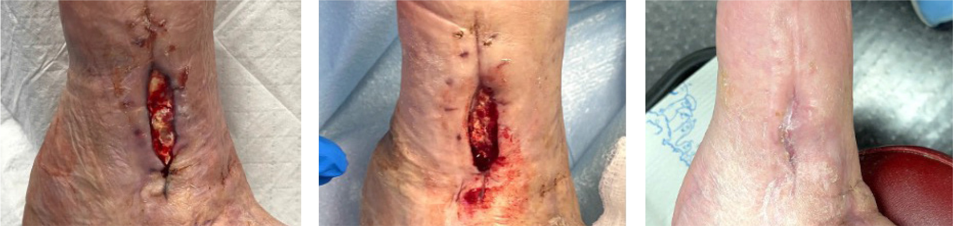
Sequential images shown in Figure 1 document the trajectory of wound healing from SV-1, TV-1, and HCV in a patient assigned to the DLAG with SOC treatment arm.
FIGURE 1 Digital images from SV-1, TV-1, and HCV (left to right), DLAG with SOC treatment arm. The patient gave consent for the publication of images.
SOC patients were offered a separate rescue trial if they failed to heal. The long-term durability trials are separate from the main trial.
Discussion
Interim analysis included a data lock on the electronic data capture (EDC) system and quality assurance review prior to data analysis. The purpose of this interim analysis is to determine balance across treatment groups and comparison to current standard of care for the primary endpoint and PAR. Patients were stratified by wound area and wound age. There is no significant difference between strata across all treatment groups, therefore, the randomization scheme achieved a balanced baseline. Additional analysis by the strati-fication group is planned for the final analysis.
For the primary endpoint, DLACG with SOC was statistically significant in the ITT population, however, was not statistically significant in the PP population. The small differences in sample size between populations may influence the results of the Chi-squared test, and additional enrollment will occur until the planned sample size is met for all treatment groups.
Percent area reduction provides insight into the closure rates by treatment group. In the ITT population, all treatment groups achieved a higher mean area reduction than SOC, while in the PP population DLACG with SOC and FLAG with SOC achieved a higher mean area reduction than SOC. This provides a promising result at interim and confirmation of the clinical trial design prior to final analysis.
Conclusion
In conclusion, the interim analysis revealed that the placental membranes products trended toward superiority over SOC. The statistical significance in the ITT population for DLACG suggests that this product is superior to SOC and once all of the ongoing patients complete, the product can be removed from the platform and replaced with a new product. The success of the placental membranes in this trial suggests that response adjusted randomization should be considered to reduce the number of patients in the SOC group.