Introduction
Around 10.5 million Medicare beneficiaries were affected by chronic wounds in 2019.1 A chronic wound is generally defined as any wound that fails to heal within a reasonable timeframe. Chronic wounds can be categorized into the following four groups: arterial, pressure, diabetic, and venous. The recurrence rate for venous leg ulcers (VLUs) is estimated to be between 60-70%.2 Despite the well-established standard of care (SOC) for chronic wounds, which includes sharp debridement, offloading, and maintaining proper moisture balance, a notable gap remains between healing outcomes and desired results in chronic wound care.
Globally, an estimated 4.5 billion individuals were affected by chronic venous disease stages C1-C6 in 2020, and approximately 0.1-0.3% of the world’s population developed a VLU.3 Despite the complex nature of VLU pathophysiology, risk factors include nonadherence with compression therapy, misdiagnosis of ulcers, obesity, and a history of deep vein thrombosis.4,5 Approximately 7% of VLUs are estimated to remain unhealed after 12 months,6 and VLUs have a recurrence rate exceeding 70% after they are closed.7 Low-income individuals, especially those from minority communities, often face difficulties in accessing advanced wound care centers.8 In 2022, the United States faced an estimated annual economic burden of over $4.9 billion USD for treating VLUs, covering costs related to healthcare practitioners, wound care products, inpatient hospitalization, medications, and compression therapy.9
Advanced wound management incorporates cellular, acellular, and matrix-like products (CAMPs), defined as ‘a broad category of biomaterials, synthetic materials, or biosynthetic matrices that support the repair or regeneration of injured tissues through various mechanisms of action’.10 CAMPs offer multiple therapeutic benefits, including protection of the wound environment, coverage of exposed deep structures, facilitation of surgical closure, and improvements in both functional outcomes and cosmetic appearance.10
The STABLECAMP clinical trial employed a modified master trial design, platform, to evaluate multiple CAMP products within a single overarching protocol. A variation of this platform design was published in 2025.11 The initial phase will include five CAMPs, three of which are included in this interim analysis. The platform design provides flexibility for addition or removal of products based on analysis of data.
Materials and methods
STABLECAMP is a multicenter, prospective, randomized controlled modified platform clinical trial designed to evaluate the efficacy of multiple cellular, acellular, and matrix-like products (CAMPs) with standard of care (SOC) compared with SOC alone for the treatment of nonhealing venous leg ulcers (VLUs) (clinicaltrials.gov #NCT06560502). The protocol initially specifies five CAMPs for evaluation; however, the adaptive nature of the platform design permits the addition of new investigational products contingent upon data analysis and protocol amendments. The interim analysis evaluated three of the five planned test products. This study was conducted at 13 SerenaGroup, Inc. or affiliated centers throughout the United States with 97 patients with nonhealing VLUs. Enrollment for this study began October 2024 and interim analysis was conducted in August 2025. The study population was drawn from patients with VLUs who were attending wound clinics.
Objectives and endpoints
The primary objective for the STABLECAMP clinical trial was to determine the between-arm difference in the proportion of subjects achieving complete closure of nonhealing diabetic foot ulcers (DFUs) and VLUs with multiple CAMPs with SOC versus SOC alone over 12 weeks. The primary endpoint was the percentage of target ulcers achieving complete wound closure in 12 weeks.
An additional important endpoint evaluated was percentage wound area reduction (PAR) from TV-1 to TV-13 measured weekly with digital photographic planimetry, using a digital imaging device and physical examination.
Diagnosis
The diagnosis of VLUs is primarily clinical and relies on a thorough patient history, comprehensive physical examination, and selective diagnostic testing. VLUs typically develop in the gaiter region of the lower leg, most commonly around the medial malleolus, and are frequently preceded by signs of chronic venous insufficiency such as edema, varicosities, lipodermatosclerosis, and hemosiderin deposition. These ulcers often present with irregular margins, a shallow wound bed, fibrinous exudate, and surrounding skin changes including hyperpigmentation or stasis dermatitis. Pain levels vary, but many patients report discomfort or aching that worsens with dependency and improves with leg elevation.
A detailed clinical history is essential to differentiate VLUs from other chronic wound types. Key elements include prior history of venous disease, episodes of deep vein thrombosis, use and adherence to compression therapy, history of ulcer recurrence, occupational or lifestyle factors involving prolonged standing, obesity, and previous wound treatments. Differential diagnoses that must be excluded include arterial ulcers, DFUs, pressure injuries, vasculitic lesions, and malignant ulcers.
All potential subjects underwent vascular screening. Ankle brachial index (ABI) was the most employed assessment. Patients with an ABI >0.7 met inclusion criteria. Values greater than 1.3 necessitated additional evaluation. In patients with incompressible, calcified arteries (common in long-standing diabetes), alternative methods such as toe-brachial index (TBI), with values ≥0.6 indicating adequate perfusion. A transcutaneous oxygen measurement (TCOM) ≥40mmHg indicating adequate perfusion also satisfied the inclusion criteria.
Vulnerable populations
Vulnerable individuals were not explicitly sought for inclusion; however, they were represented within the pool of potential participants.
Product description
This ongoing study evaluates five products: dual layer amniotic membrane allograft (DLAG), three-layer amniotic membrane allograft (TLAG), four-layer amniotic mem-brane allograft (FLAG), dual layer amnion/chorion membrane allograft (DLACG), and three-layer amnion/chorion/amnion membrane allograft (TLACG). All these products were intended for homologous use as a barrier and applied as a covering to offer protection from the surrounding environment. The products were provided sterilized in an inner peel pouch, within a non-sterile outer peel pouch, within a carton. The products are comprised of donated human tissue of which donor eligibility determinations, recovery, processing, storage, testing, and distribution are performed in accordance with 21 CFR Part 1271 and States regulations as well as AATB standards. For the interim analysis, only DLAG, FLAG, and DLACG were evaluated. Table 1 describes the features of each product.
TABLE 1 Product details
| Product name | Key features |
|---|---|
| AmnioCore | Dual layer, amniotic membrane allograft |
| Amnio Tri-Core | Three-layer, amniotic membrane allograft |
| Amnio Quad-Core | Four-layer, amniotic membrane allograft |
| AmnioCore Pro | Dual layer, amnion/chorion membrane allograft |
| AmnioCore Pro+ | Three-layer, amnion/chorion/amnion membrane allograft |
Subject characteristics
Patients who suffer from nonhealing VLUs were recruited for this study from participating wound clinics. Once patients agreed to adhere to the study schedule and read and signed the IRB approved Informed Consent Form, screening was conducted to determine whether subjects were eligible based on inclusion and exclusion criteria, listed in Table 2.
TABLE 2 Inclusion and exclusion criteria
| Inclusion criteria | Exclusion criteria |
|---|---|
|
|
Study procedures
Participants underwent a structured sequence of clinical visits including screening, treatment, healing confirmation, and follow-up phases to ensure accurate eligibility assessment, standardized wound care, consistent intervention delivery, and reliable endpoint determination. Subjects were evaluated weekly (±3 days) over a 12-week treatment period, with any additional dressing changes recorded as unscheduled visits and abbreviated assessments performed when needed.
Participants who did not meet eligibility criteria at initial screening but were subsequently determined eligible were re-consented and assigned a new screening number. Up to three screening attempts were allowed, and those who subsequently met all inclusion and no exclusion criteria were enrolled.
Screening visits
At the screening visit, conducted approximately 14 days prior to enrollment, informed consent was obtained, followed by a review of medical history to assess eligibility based on inclusion and exclusion criteria. Demographic data (including height, weight, BMI, sex, and ethnicity), medical and medication histories, and current use of non-steroidal anti-inflammatory drugs (NSAIDs) and opioids were recorded. A vascular screening test was performed unless recent results (≤3 months) were available. Vital signs were measured, and a general physical examination was conducted. Additional assessments included the Mini Nutritional Assessment (MNA), HbA1c testing (unless available within 3 months), clinical, etiologic, anatomic, and pathophysiologic (CEAP) classification, Fitzpatrick skin type, pain intensity via a visual analogue scale (VAS), and detailed wound characterization (granulation tissue, nonviable tissue, depth, exudate, and peri-wound skin).
The CEAP classification is an internationally accepted system for the standardized assessment of chronic venous disorders (CVD). Clinical (C) describes the visible or palpable clinical signs, ranging from no visible disease (C0) to active venous ulceration (C6). Etiologic (E) identifies the cause as congenital, primary, or secondary (post-thrombotic). Anatomic (A) specifies the location of the venous abnormality within the superficial, deep, or perforating systems. Pathophysiologic (P) denotes the underlying mechanism—reflux, obstruction, or both. This structured approach facilitates consistent diagnosis, comparison of clinical outcomes, and communication among clinicians and researchers studying venous disease.
Historical wound measurements from two weeks prior to screening were collected; a reduction in wound size of >20% during this historical run-in period resulted in screen failure.
During the two-week active screening phase, SOC wound management was provided, consisting of cleansing with normal sterile saline (NSS), sharp debridement, post-debridement ulcer photography and measurement using a digital imaging device, and application of calcium alginate or foam dressing. Other dressings were not permitted without permission from the medical monitor.
Treatment visits
At the enrollment/randomization visit (treatment visit (TV) 1; Day 0), eligibility was re-confirmed, adverse events and medication changes were reviewed, and a symptom-directed physical examination was performed. The percentage area reduction (PAR) from the screening period was assessed to confirm it remained <20%. Vital signs, wound characteristics, VAS pain scores, Forgotten Wound Score (FWS), and the Wound Quality of Life (wQOL) questionnaire were completed. VLU participants also underwent Functional Ambulatory Category Scale (FACS) assessment. Eligible participants were randomized to receive either CAMPs with SOC or SOC alone. Wounds were cleansed with NSS, debrided, photographed, and measured using a digital imaging device before treatment. Dressings were applied as per protocol, with optional absorptive dressings for highly exudative wounds upon medical monitor approval.
Participants returned weekly (TV-2 to TV-12) for safety and efficacy monitoring, including review of adverse events, medication changes, vital signs, wound assessment, pain scoring, and questionnaire administration (FWS and wQOL at TV-4, TV-8, and TV-12). Wound cleansing, debridement, measurement, and treatment per randomization arm were repeated at each visit.
At the final treatment visit (TV-13) or earlier if wound closure occurred, adverse events, medication changes, pain, and quality-of-life measures (FWS, wQOL) were recorded. Participants also underwent FACS assessment. For ulcers that had not yet healed, wound characteristics were documented, and follow-up care was arranged. Healing was confirmed at a dedicated visit 14±3 days after the first observation of complete re-epithelialization without drainage. This visit included adverse event review, medication update, pain assessment, investigator confirmation of closure, ulcer photography and measurement, and independent blinded verification. Table 3 shows the study schedule.
TABLE 3 Study schedule
| SV | TV-1 | TV-2, TV-3 | TV-4 | TV-5, TV- 6, TV-7 | TV-8 | TV-9, TV- 10, TV-11 | TV-12 | TV-13 | CCV | |
|---|---|---|---|---|---|---|---|---|---|---|
| Window period | -14 | Day 0 | Week 1,week 2 | Week 3 | Week 4,Week 5,Week 6 | Week 7 | Week 8Week 9Week 10 | Week 11 | Week 12 | 14+ |
| Record medical history and demographic information | X | |||||||||
| Assessment of eligibility | X | X | ||||||||
| Sign informed consent form | X | |||||||||
| Vascular screening test | ||||||||||
| Physical exam | X | X | ||||||||
| Mini Nutrition Assessment | X | |||||||||
| HbA1c | X | |||||||||
| CEAP Classification | X | |||||||||
| Fitzpatrick Scale | X | |||||||||
| Historical measurement | X | |||||||||
| Randomization | X | |||||||||
| Assessment for AE and SAE | X | X | X | X | X | X | X | X | X | |
| Review medication for changes | X | X | X | X | X | X | X | X | X | |
| Vital signs | X | X | X | X | X | X | X | X | ||
| Wound assessment | X | X | X | X | X | X | X | X | X | X |
| Pain assessment (VAS) | X | X | X | X | X | X | X | X | X | X |
| wQOL | X | X | X | X | X | |||||
| FWS | X | X | X | X | X | |||||
| FACS | X | X | ||||||||
| Study ulcer cleaning, debridement (if applicable) | X | X | X | X | X | X | X | X | X | |
| Study ulcer area with digital images | X | X | X | X | X | X | X | X | X | X |
| Treatment based on randomization | X | X | X | X | X | X | X | |||
| Apply dressing | X | X | X | X | X | X | X | X | X |
Study exit
Early withdrawals underwent procedures equivalent to the final visit when possible. Unscheduled visits were conducted as needed for adverse event evaluation, medication review, and dressing changes. At study exit, participants with wounds that had not yet healed were transitioned back to physician-directed SOC. Independent healing confirmation was performed by two blinded wound care specialists reviewing de-identified digital images from closure and confirmation visits. Disagreements between reviewers were resolved in favor of the assessment aligning with the principal investigator’s determination.
Subject withdrawal
All participants had the right to withdraw from the study at any time during the treatment period without prejudice. The completion status of each participant’s involvement in the clinical trial was documented. In the event that study treatment or protocol-required observations were discontinued for any participant, the reason(s) for discontinuation were recorded. The investigator had the authority to withdraw a participant from the study at any time if deemed medically necessary. Whenever feasible, the reason for withdrawal or early termination was documented.
A participant was classified as lost to follow-up if they could not be reached after five telephone contact attempts and three written communications.
Subject compensation
Participants received a nominal compensation of $50 USD upon completion of each study visit. This compensation was intended to offset expenses associated with participation, including travel, parking, and the additional time required for study-specific procedures and data collection.
Results
A total of 110 VLU patients from 13 sites were enrolled in the interim analysis. 24 patients received SOC alone, 20 patients received DLAG with SOC, 6 patients received DLACG with SOC, and 17 patients received FLAG with SOC. Additionally, 9 patients from all treatment groups are considered ongoing at the time of interim analysis. Overall, 17 patients were discontinued during the study and 17 were excluded during screening. Summary statistics on demographic variables are provided in Table 4 and Table 5.
TABLE 4 Demographic summary statistics by treatment group
| Variable | Standard of care | DLAG | DLACG | FLAG | P-value |
|---|---|---|---|---|---|
| Age (Years) | |||||
| N (Mean) | 23 (69.7) | 20 (68.3) | 6 (65.3) | 16 (69.9) | 0.869 |
| Sex, N (%) | |||||
| Male | 11 (47.8) | 9 (45.0) | 3 (50.0) | 8 (50.0) | 0.959 |
| Female | 12 (52.1) | 11 (55.0) | 3 (50.0) | 8 (50.0) | |
| Fitzpatrick Scale, N (%) | |||||
| Type I | 5 (22.7) | 5 (25.0) | 1 (16.6) | 5 (31.2) | 0.895 |
| Type II | 4 (18.1) | 7 (35.0) | 2 (33.3) | 5 (31.2) | |
| Type III | 6 (27.2) | 3 (15.0) | 1 (16.6) | 1 (6.2) | |
| Type IV | 1 (4.5) | 2 (10.0) | 0 (0) | 3 (18.7) | |
| Type V | 4 (18.1) | 2 (10.0) | 1 (16.6) | 1 (6.2) | |
| Type VI | 2 (9.0) | 1 (5.0) | 1 (16.6) | 1 (6.2) | |
| Race, N (%) | |||||
| American Indian/Alaskan Native | 0 (0) | 0 (0) | 0 (0) | 0 (0) | 0.594 |
| Asian | 0 (0) | 1 (5.0) | 0 (0) | 0 (0) | |
| Black | 8 (34.7) | 5 (25.0) | 3 (50.0) | 3 (18.7) | |
| Pacific Islander | 0 (0) | 0 (0) | 0 (0) | 0 (0) | |
| White | 10 (43.4) | 13 (65.0) | 3 (50.0) | 11 (68.7) | |
| Other | 5 (21.7) | 1 (5) | 0 (0) | 2 (12.5) | |
| Current tobacco use, N (%) | |||||
| Yes | 5 (21.7) | 5 (25.0) | 1 (16.6) | 1 (6.2) | 0.716 |
| No | 18 (78.2) | 15 (75.0) | 5 (83.3) | 15 (93.7) | |
TABLE 5 Stratification summary statistics
| Variable | Standard of care (n = 24) | DLAG (n = 20) | DLACG (n = 6) | FLAG (n = 17) | P-value |
|---|---|---|---|---|---|
| Wound area, N (%) | |||||
| Less than 2cm2 | 3 (12.5) | 4 (20.0) | 1 (16.6) | 3 (17.6) | 0.354 |
| Between 2cm2 and 3cm2 | 4 (16.6) | 5 (25.0) | 1 (16.6) | 5 (29.4) | |
| Greater than 3cm2 | 17 (70.8) | 11 (55.0) | 4 (66.6) | 9 (52.9) | |
| Wound age, N (%) | |||||
| Wound >60 days | 6 (25.0) | 2 (10.0) | 1 (16.6) | 2 (11.7) | 0.426 |
| Wound <60 days | 18 (75.0) | 18 (90.0) | 5 (83.3) | `15 (88.2) | |
No statistically significant differences were observed across treatment groups (all p>0.05), suggesting that randomization achieved adequate baseline balance. Wound area and wound age were used as stratification factors in the trial design, and at this interim analysis, they are summarized descriptively to assess balance. The reported p-values are exploratory checks of randomization balance and were not used to adjust the interim analysis endpoints.
The primary endpoint was assessed for the interim analysis. The primary endpoint is the percentage of target ulcers achieving complete wound closure in 12 weeks. The intent-to-treat (ITT) and per protocol (PP) populations were analyzed.
In the ITT population, DLAG with SOC arm achieved a 50% closure rate versus 16.7% with SOC alone, an 33.3% absolute gain that was statistically significant (n=20, 95% CI 5.6% to 55.8%, p=0.018, α=0.05). The DLACG with SOC arm achieved a 16.7% closure rate versus 16.7% with SOC alone, with no absolute gain that was not statistically significant (n=6, 95% CI -23.6% to 40.9%, p=1, α=0.05).The FLAG with SOC arm achieved a 11.8% closure rate versus 16.7% with SOC alone, a 4.9% absolute gain that was not statistically significant (n=17, 95% CI -25.9% to 19.8%, p=0.662, α=0.05).
Among the PP population, DLAG with SOC arm achieved a 66.7% closure rate versus 44.4% with SOC alone, a 22.3% absolute gain that was not statistically significant (n=12, 95% CI -17.7% to 54.4%, p=0.309, α=0.05). The DLACG with SOC arm achieved a 33.3% closure rate versus 44.4% with SOC alone, a 11.1% absolute gain that was not statistically significant (n=3, 95% CI -50.8% to 41.4%, p=0.735, α=0.05). The FLAG with SOC arm achieved a 50% closure rate versus 44.4% with SOC alone, a 5.6% absolute gain that was not statistically significant (n=4, 95% CI -39.8% to 48.9%, p=0.853, α=0.05).
Additionally, the percent area reduction (PAR) from TV-1 to TV-13 was measured weekly with digital photographic planimetry using an imaging device, and physical examination were analyzed.
Within each arm, any individual PAR value falling below Q1 – 1.5*IQR or above Q3 + 1.5*IQR was flagged and excluded. For ITT, DLAG with SOC outperformed SOC on both average and median wound-area reduction, with summary statistics for each treatment group (without outliers) reported in Table 6.
TABLE 6 Percent Area Reduction (PAR) summary statistics without outliers for ITT
| Treatment arm | N | Mean | Standard deviation | Median | IQR |
|---|---|---|---|---|---|
| Standard of Care | 24 | 40.93 | 44.42 | 39.56 | 81.86 |
| DLAG with SOC | 20 | 49.60 | 67.30 | 96.90 | 84.46 |
| DLACG with SOC | 6 | 24.35 | 68.80 | 44.42 | 87.40 |
| FLAG with SOC | 16 | 4.58 | 89.39 | 32.56 | 75.73 |
For PP, DLAG with SOC outperformed SOC on both average and median wound-area reduction, with summary statistics for each treatment group (without outliers) re-ported in Table 7.
TABLE 7 Percent Area Reduction (PAR) summary statistics without outliers for PP
| Treatment arm | N | Mean | Standard deviation | Median | IQR |
|---|---|---|---|---|---|
| Standard of Care | 9 | 66.37 | 42.60 | 81.08 | 55.62 |
| DLAG with SOC | 12 | 71.88 | 65.98 | 100.0 | 0 |
| DLACG with SOC | 3 | 31.01 | 97.08 | 73.02 | 90.0 |
| FLAG with SOC | 4 | 16.18 | 96.98 | 20 | 163.82 |
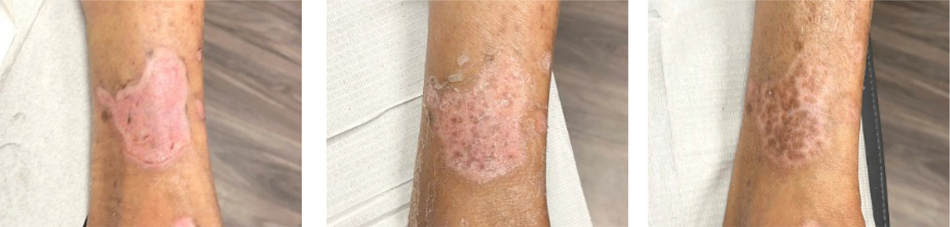
Sequential images shown in Figure 1 document the trajectory of wound healing from SV-1, TV-1, and HCV in a patient assigned to the DLAG with SOC treatment arm.
FIGURE 1 Digital images from SV-1, TV-1, and HCV (left to right), DLAG with SOC treatment arm. Patients gave consent for publication of images.
Discussion
Interim analysis included a data lock on the electronic data capture (EDC) system and quality assurance review prior to data analysis. The purpose of this interim analysis is to determine balance across treatment groups and comparison to current standard of care for the primary endpoint and PAR. Patients were stratified by wound area and wound age. There is no significant difference between strata across all treatment groups, therefore, the randomization scheme achieved a balanced baseline. Additional analysis by the stratification group is planned for the final analysis.
For the primary endpoint, DLAG with SOC was statistically significant in the ITT and PP population. The small differences in sample size between populations may influence the results of the Chi-squared test, and additional enrollment will occur until the planned sample size is met for all treatment groups.
Percent area reduction provides insight into the closure rates by treatment group. In the ITT and PP population, DLAG with SOC achieved a higher mean area reduction than SOC. This provides promising results at interim and confirmation of the clinical trial design prior to final analysis.
The limitation of this interim analysis, like most interim looks, is the small number of patients. The primary purpose of interim analysis is to determine if the treatment arms are trending toward superiority over SOC alone. We can say with confidence that the treatment arms are outperforming SOC.
Conclusion
In conclusion, the interim analysis revealed that the placental membranes products trended towards superiority over SOC. The statistical significance in the ITT population for DLACG suggests that this product is superior to SOC and once all of the ongoing patients complete their treatment, the product can be removed from the platform and replaced with a new placental membrane. The success of the placental membranes in this trial suggests that response-adjusted randomization should be considered to reduce the number of patients in the SOC group. Finally, this analysis demonstrates the power of the modified master trial design in simultaneously evaluating multiple CAMP products.