Introduction
Chronic wounds remain a key health concern, affecting 1–2% of the global population,1 and posing a substantial burden on healthcare systems.2-6 Their chronicity is influenced by complex patient- and wound-level factors such as bacterial infection, tissue hypoxia, metabolic disease, and impaired perfusion that can impede tissue healing.7-9 Despite the complexity of these conditions, clinical wound assessment commonly relies largely on subjective visual inspection and manual measurement, which vary by observer and delay recognition of poor treatment responses and early signs of infection.10-13 Some of these early signs, such as bacterial burden, tissue hypoxia, and inflammation may not be visible or palpable during routine clinical examination, potentially delaying their recognition and appropriate intervention.14,15 Conventional methods (visual assessment and manual wound measurements) have provided limited information to clinicians regarding the underlying physiological status of wounds at the point-of-care (POC).
The recent advances in optical and digital imaging technologies have allowed clinicians to visualize microbial burden, tissue oxygenation, and perfusion at the bedside during wound assessment.16-19 These imaging systems may provide objective and quantitative information that can complement traditional clinical assessment and guide more targeted interventions.12,16 Fluorescence imaging has been established as a POC device for real-time elevated bacterial detection in chronic wounds.20 The MolecuLightDX+ utilizes safe violet light (405 nm) excitation to visualize endogenous bacterial fluorescence, enabling detection of clinically significant bacterial loads (>104 CFU/g) at POC.20-22 By identifying areas of elevated bacterial burden, fluorescence imaging may support targeted cleansing and debridement and provide additional information to inform antimicrobial or antibiotic decisions, which have shown to improve healing and minimize the risk of wound-related complications.20,23-25
Thermal imaging has been used in clinical diagnostics, as a non-invasive, contact-free method for detecting physiological disturbances through surface temperature variations.26,27 In wound care, thermal imaging has been used to monitor healing, detect signs of complications, and predict outcomes in diabetic foot ulcers, pressure injuries, and surgical wounds.28 Thermal imaging measures skin surface temperature gradients that may be associated with inflammatory, infection or ischemic processes.28-31 Thermal imaging has also been explored as a non-invasive tool for identifying deeper infections, such as osteomyelitis, in patients with diabetic foot ulcers, as elevated skin temperatures extending beyond the wound can indicate underlying inflammatory activity.32,33 By translating infrared radiation into color maps, thermography has helped in the detection of subtle temperature variations that may be associated with inflammation, tissue necrosis, and healing progression before observable clinical signs and symptoms emerge.27,28,34-36 Thermography has shown to be sensitive to ambient environmental conditions and subject to calibration variability, both of which can affect measurement accuracy;37 however, recent advances have enabled the development of portable, POC systems capable of real-time temperature assessment.28,38 The combined use of fluorescence, and thermal imaging may provide additional information regarding bacterial burden and underlying tissue physiology during clinical wound assessment. The potential value of combining these modalities in routine practice, particularly their influence on clinical decision making remains incompletely described.28
The aim of this case series is to demonstrate how clinicians utilize multimodal fluorescence and thermal imaging in the management of mixed chronic wounds. We present five cases to illustrate how information on thermal distribution and bacterial burden can support real-time clinical decision making, optimize cleansing and debridement, and improve monitoring of tissue recovery.
Methods
Case collection and setting
This descriptive case series includes patients who were observed at and received care from four wound care centers; the Madigan Army Medical Center (Tacoma, WA, USA), the Northwell Comprehensive Wound Health and Hyperbaric Center (Lake Success, NY, USA), the Scarborough Health Network (Toronto, ON, Canada), and the Judy Dan Research and Treatment Centre (North York, ON, Canada). The wound imaging device has been used as a part of the routine clinical practice at the participating centers. Cases for this descriptive case series were selected based on having sufficient clinical documentation to illustrate the clinical application of multimodal imaging for chronic wound management in routine care, without a predefined study cohort. Patients presented in this case series were assessed between November 2023 and January 2025. Patient data, wound assessment results, and treatment plans were recorded and deidentified to protect patient confidentiality. Written, informed consent was obtained from all participants, including for the publication of photographs or images. Ethics approval was not required for this noninterventional, observational case series, which was performed in accordance with the Helsinki Declaration of 1964 and its later amendments.
Wound imaging device and procedures
MolecuLightDX+ (MolecuLight Inc., Toronto, Ontario, Canada) is a handheld POC fluorescence imaging device that is used in wound care settings to detect elevated bacterial loads (>104 CFU/g) in wounds, relative temperature differentials and perform digital wound measurements.
All four clinicians participating in the study received standardized training on device operation and image interpretation and had several years of hands-on experience with the device in their clinical practice. Fluorescence imaging was conducted in a darkened environment and from an optimal distance of approximately 8–12 cm. During fluorescence imaging, the device emits safe violet light at a wavelength of 405 nm resulting in porphyrin-producing bacteria generating red fluorescence, Pseudomonas species producing cyan signals from pyoverdines, and collagen and elastin within tissues emit green fluorescence, all captured in the images. Fluorescence images were interpreted by the treating clinician and a positive fluorescence result was defined as the detection of red or cyan signals within the wound bed or surrounding tissue.
The MolecuLight thermal module was used to capture and analyze temperature distributions across the wound bed and periwound tissue. Thermal images of the wound were captured at appropriate distances to include both contralateral limbs or corresponding anatomical area as a reference comparator. Temperature differences (ΔT °C) were calculated relative to adjacent intact skin, periwound tissue or the contralateral limb depending on the clinical presentation and anatomical location of the wound. The device also enabled co-registered imaging of the wound, where standard, thermal and fluorescence images were taken almost simultaneously. The module generates a color-coded thermal scale ranging from –6 °C to +5 °C, relative to a user-selected reference point on adjacent intact skin (green). Cooler regions (light to dark purple) reflect temperatures lower than the reference point and may be associated with reduced local perfusion, undermining, or devitalized tissue. Warmer regions (yellow to red) appear when temperatures exceed the reference point and may indicate areas of localized inflammation or infection. Temperature differences allow interpretation of spatial thermal gradients in the wound and surrounding tissue. Thermal images were taken approximately 5 minutes after wound undressing to allow for acclimatization but prior to cleansing and debridement. Clinical interpretation of the thermal and fluorescence imaging results and the treatment, including additional debridement or change in wound management plan, was at the discretion of the treating clinician.
Results
Five cases are presented to illustrate the use of multimodal thermal and fluorescence imaging across a range of chronic wound presentations. Each case demonstrates how the combined use of thermal and fluorescence imaging provided additional objective information that supports standard clinical assessment. Thermography findings were suggestive of areas of ischemia, inflammation, undermining, and tunneling, while fluorescence imaging identified regions of elevated bacterial burden. These imaging findings were interpreted alongside clinical assessment and incorporated into patient management by the treating clinician, which led to informed clinical interventions.
Case 1: Detecting unseen vascular impairment
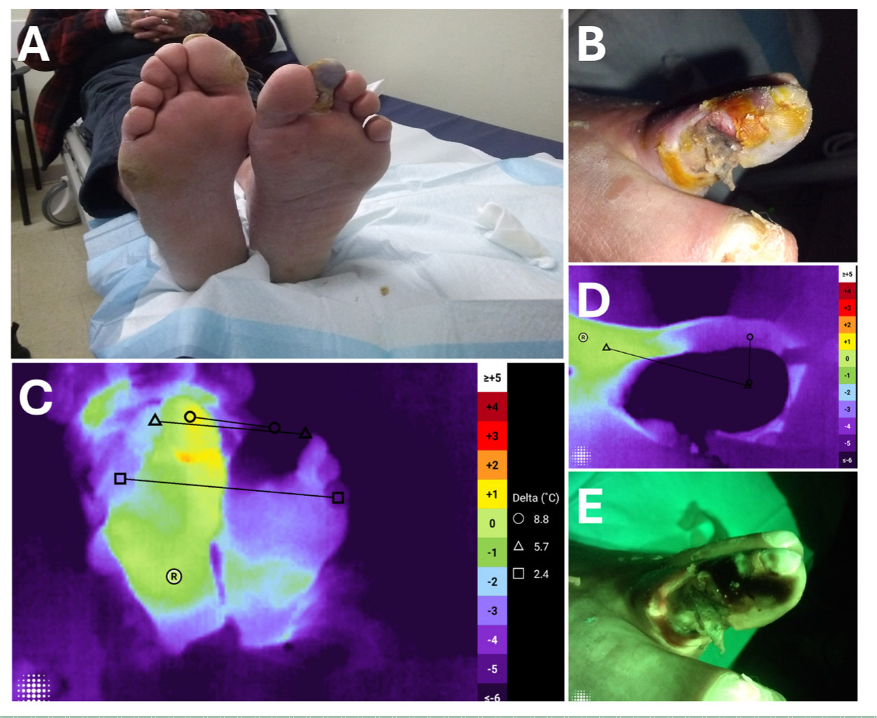
A 60-year-old male with diabetes, Crohn’s disease, and a long-term smoking history presented with a painful, chronic ulcer on the left foot that had persisted for 6 weeks. Standard wound assessment (Figure 1A and B) revealed cyanosis of the second left toe, consistent with ischemia, while the first toe appeared clinically unremarkable. Additional assessment with thermal and fluorescence imaging was conducted to observe any thermal changes. By comparing relative temperature difference between the contralateral limbs, thermal imaging (Figures 1C and D) demonstrated a ΔT that may be suggestive of ischemia; the second toe was 5.7°C cooler than the contralateral foot. Thermal imaging also revealed a cooler temperature in the great toe compared with the contralateral foot (ΔT = -8.8°C), which may be suggestive of blood flow impairment that was not apparent during the initial clinical assessment. These findings raised concern for more extensive ischemia than initially suspected, and prompted immediate referral for further vascular assessment focusing on the first toe. Fluorescence imaging (Figure 1E) showed no elevated bacterial signal. Follow-up information shows that the patient later underwent vascular assessment, followed by angiography and angioplasty to restore perfusion in the dorsalis pedis artery. In this case, thermal imaging identified areas of relative temperature differences later confirmed as impaired perfusion that were not apparent on standard examination, enabling earlier vascular assessment and intervention.
FIGURE 1 Detection of unseen vascular impairment using thermal and fluorescence imaging. (A–B) Standard clinical images showing cyanosis of the second toe, while the first toe appears intact. (C–D) Thermal image demonstrating a significant temperature difference (second toe 5.7°C cooler and first toe 8.8°C cooler than contralateral limb), consistent with widespread ischemia. (E) Fluorescence image showed no elevated bacterial signal.
Case 2: Detecting undermining and devitalized tissue
A 58-year-old female presented with an unstageable pressure ulcer on the plantar surface of right foot. Standard wound assessment (Figure 2A) did not show clinical signs and symptoms of infection, however the edges of the wound did appear macerated. Thermal imaging (Figure 2B) revealed cooler zones (ΔT = -5.8°C) along the lower right quadrant of the wound compared with the periwound, suggesting possible undermining. A localized cool zone (ΔT = -1.7°C) suggestive of devitalized tissue was also observed in the top left quadrant of the wound. This finding led to probing of the wound, and identified a significant pocket of undermining in the bottom right quadrant consistent with the decreased temperature in that area. This region was selected for debridement by the clinician. Following debridement, fluorescence imaging (Figure 2C) showed a localized blush-red signal indicating elevated bacterial burden, which was not clinically apparent on visual inspection alone.
FIGURE 2 Thermal and fluorescence imaging reveal undermining and devitalized tissue in a pressure ulcer. (A) Standard image of the pressure ulcer on plantar surface of right foot did not show any clinical signs and symptoms of infection, although the wound edges appeared macerated. (B) Thermal map highlighting cooler zones along the lower-right wound edge (ΔT = -5.8°C) and upper-left quadrant (ΔT = -1.7°C), indicating undermining and devitalized tissue. (C) Fluorescence image showing localized blush-red signal at the wound margin.
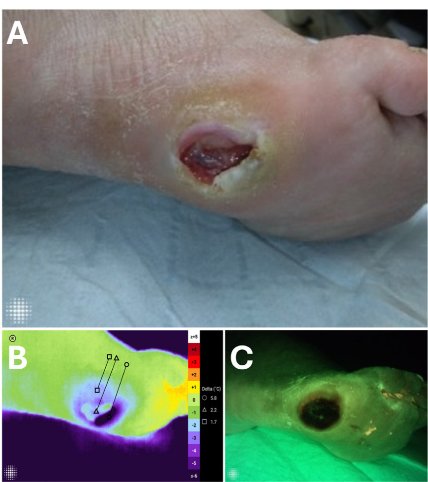
Together, thermal and fluorescence imaging identified areas suggestive of undermining and elevated bacterial burden that were not apparent on visual assessment, prompting further debridement by the treating clinician.
Case 3: Detecting hidden tunneling
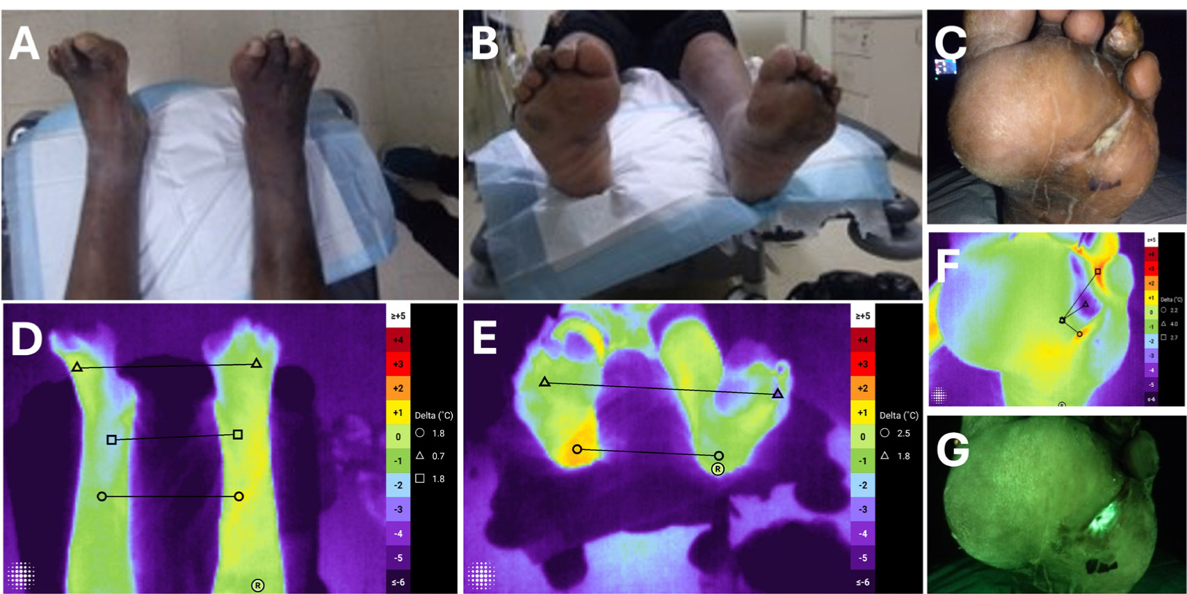
A 38-year-old male with type 2 diabetes presented with a chronic plantar wound on the left foot. Standard wound assessment (Figures 3A-C) did not show clinical signs and symptoms of infection. Thermal imaging of both limbs (Figures 3D and E) showed a small ΔT of -1.8°C. Localized thermal imaging (Figure 3F) identified a 4.0°C cooler zone within the wound bed, which was not immediately apparent during clinical examination of the foot. This ΔT in the wound bed indicated probable presence of a deep tunneling tract, which was confirmed by probing of the wound. Fluorescence imaging (Figure 3G) revealed cyan signal within the wound bed, suggestive of Pseudomonas aeruginosa presence. Subsequent tissue sampling and microbiological analysis confirmed the presence of P. aeruginosa. Together, fluorescence imaging and thermal analysis helped the clinician’s decision to pack the wound tunnel and apply Hydrofera Blue and a dry dressing to the wound to address the Pseudomonas presence.
FIGURE 3 Multimodal detection of hidden tunneling and bacterial contamination in a diabetic foot wound. (A–B) Standard photographs of both feet demonstrating intact right foot and a shallow left plantar wound. (C) Close-up standard image of the left plantar ulcer showing well-defined margins and minimal exudate. (D–E) Thermal images of both feet showed a small ΔT of -1.8°C. (F) High-resolution thermal image of the wound revealing a localized cooler tract consistent with hidden tunneling not evident on visual inspection. (G) Fluorescence image displaying cyan signal within the wound bed indicative of Pseudomonas aeruginosa. Abbreviations: ΔT, temperature difference.
Case 4: Supporting infection detection and treatment response
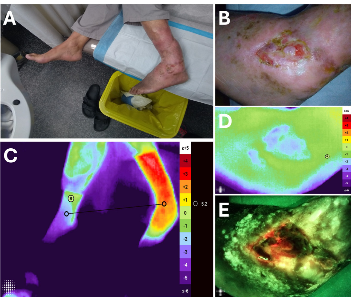
A 62-year-old male presented with a chronic traumatic wound (Figure 4A and B) on the right ankle that had persisted for over 35 years despite multiple reconstructive surgeries. The standard wound assessment showed localized periwound erythema and swelling. Thermal imaging (Figure 4C and D) showed a 5.2°C warmer temperature over the affected ankle relative to the contralateral ankle. Fluorescence imaging (Figure 4E) showed red signals along the cooler wound edges, that may indicate high bacterial presence and possible devitalized tissue. Together, these findings raised suspicion of infection, which was confirmed by microbiological analysis of the collected tissue demonstrating the presence of Staphylococcus aureus. Systemic antibiotic therapy was initiated, and a silver dressing was applied on the wound. Thermal and fluorescence imaging was also used to monitor treatment response, tracking the presence of bacterial fluorescence and a reduction in the relative temperature difference between limbs. In this case, thermal and fluorescence imaging identified findings suggestive of infection that were subsequently microbiologically confirmed and helped clinicians in treatment planning and follow-up assessment.
FIGURE 4 Infection detection and response monitoring in a chronic traumatic ankle wound. (A-B) Standard images of foot and wound. (C-D) Thermal image demonstrating a 5.2°C temperature elevation over the affected ankle relative to the contralateral side, suggestive of localized inflammation or possible infection. (E) Fluorescence image revealing intense red signals at cooler peripheral zones, corresponding to devitalized tissue with bacterial burden.
Case 5: Detecting infection in a pressure injury in a patient with Fitzpatrick VI skin type
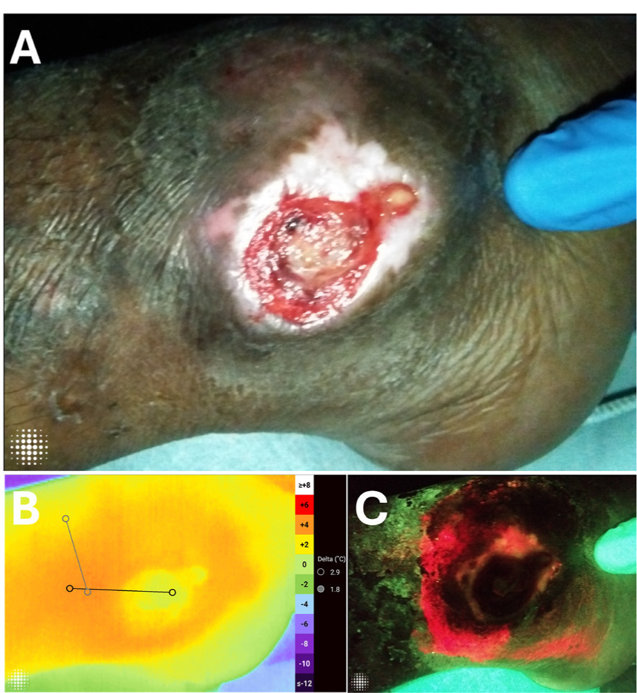
A 24-year-old male patient with paraplegia receiving home-nursing support presented with chronic pressure ulcers over the right ankle region (Figure 5A). The patient had Fitzpatrick skin type VI, in which visual detection of erythema and inflammation is often challenging,39 limiting the reliability of visual assessment alone. The wounds were evaluated using fluorescence imaging to assess the bacterial burden. Thermal imaging (Figure 5B) demonstrated elevated temperature in the periwound region compared with the wound bed (ΔT ≈ 3 °C), as well as a smaller temperature difference between the periwound tissue and more distant surrounding tissue (ΔT ≈ 2 °C). These temperature gradients suggested potential inflammation and infection around the wound area. Fluorescence imaging (Figure 5C) revealed a red signal within the wound and periwound region, indicating bacterial burden. Based on the positive fluorescence findings and increased temperatures in the periwound, the wound was swabbed for microbiology to identify the causative pathogens and bacterial loads, later confirming the fluorescence imaging and informing appropriate antibiotic selection.
FIGURE 5 Identification of infection and inflammatory hotspot in a chronic right-ankle pressure ulcer. (A) Standard image of the pressure ulcer. (B) Thermal image showing a localized warmth at the wound; ΔT ≈ 3°C between the wound bed and periwound, and ΔT of ≈ 2°C between the periwound and more distant surrounding tissue. (C) Fluorescence image showing the red signal within the wound and periwound area, consistent with elevated bacterial load. Abbreviations: ΔT, temperature difference.
Discussion
This 5-case observational series describes the use of thermal and fluorescence imaging that provided additional clinically valuable insights into wound pathophysiology, specifically, the interplay between perfusion, inflammation and bacterial burden across a variety of chronic wounds. Thermal imaging identified ΔT values, revealing areas suggestive of ischemia, inflammation, undermining or tunneling that were not visible on clinical assessment. Fluorescence imaging revealed areas of localized bacterial burden across cases, which supported timely interventions such as debridement and antimicrobial or antibiotic therapy. Collectively, these findings show how temperature mapping and bacterial fluorescence can provide complementary information for wound assessment.
The present case series demonstrates how multimodal thermal and fluorescence imaging may support clinical decision-making by providing objective physiological and microbiological information not available through routine wound assessment. In one case, thermal imaging identified temperature differences that were not clinically evident. This observation aligns with findings by Lin et al,30 who showed that cooler surface temperatures on the foot compared with adjacent healthy tissue (i.e., reference point on the foot) may indicate reduced blood supply. Similarly, Koerner et al observed that a ΔT of approximately 4°C to 5°C may be associated with ischemic tissue compared to healthy skin.40 These findings supported clinically relevant decisions, including vascular assessment and referral to a vascular surgeon.
In another case, cooler thermal zones along the wound edge were observed, consistent with the findings of Kanazawa et al, who reported that lower wound edge temperatures may correspond to areas of potential undermining.41 This may be suggestive of devitalized or necrotic tissue, consistent with previous reports showing underlying tissue changes or altered perfusion.29,42 A localized cooler region was also observed with a tunneling tract, consistent with prior reports suggesting that focal hypothermic zones may indicate hidden tunneling or limited microvascular perfusion.29,41 In chronic traumatic and pressure injuries, thermal differences, manifested as elevated limb temperature or focal periwound hotspots, were observed, consistent with previous reports that surface temperature elevation can serve as an early indicator of infection.13,34,43
Thermal and fluorescence imaging provide complementary information related to perfusion (measured indirectly using thermal imaging), microbiology (fluorescence imaging) and morphology (standard color imaging). In addition to these imaging modalities, the MolecuLightDX+ device used in this case series provides digital wound measurement capabilities, including automatic measurement of the deepest visual wound depth, that helps in longitudinal monitoring of changes in wound size over time.
This case series illustrates real-world applications of multimodal imaging in the management of a variety of wound types. Interpretation of imaging results was performed by clinicians who had received standardized training and had experience in device use, as well as fluorescence and thermal imaging. As a retrospective descriptive case series, the findings are subject to selection bias as the cases presented were chosen to illustrate the clinical application of multimodal imaging. The study was intended to provide descriptive clinical observations rather than evaluate the diagnostic performance or clinical outcomes, therefore no blinded assessments or comparator groups were included. The small sample size limits the generalizability of the findings. Clinical interpretation of imaging findings and subsequent management decisions remained at the discretion of the treating clinician. Additional limitations include environmental factors, such as ambient room temperature and patient acclimatization time, which may affect thermal imaging, as well as the need for darkened conditions during fluorescence imaging. Interpretation of thermal and fluorescence imaging findings requires appropriate training and clinical experience. Fluorescent signals indicate the presence of elevated bacterial burden rather than confirmation of infection and should be considered alongside clinical findings.
Future prospective studies are needed to validate the findings of this case series, evaluate their impact on clinical outcomes, and assess the cost-effectiveness of multimodal imaging in chronic wound care.
Conclusions
This case series describes the clinical application of a combined thermal and fluorescence approach in chronic wound assessment. Together with the digital wound measurement module, the device can provide a multimodal, three-in-one solution for chronic wound assessment and for guiding the most appropriate course of therapy. Assessing fluorescence and thermal modes may provide additional information on perfusion changes, undermining or tunneling, inflammation and bacterial burden at the point-of-care supporting clinical assessment and management decisions. The case series shows that thermal imaging provides a useful addition to multimodal wound assessment by providing visual and quantitative information regarding temperature variation within the wound and surrounding tissue, and may be helpful in monitoring healing trajectories.