Introduction
Calciphylaxis, also known as calcific uremic arteriolopathy, is a rare and often fatal condition characterized by calcification and thrombosis of small- to medium-sized arterioles, leading to tissue ischemia and necrosis.1 It occurs most commonly in patients with end-stage renal disease (ESRD) receiving dialysis, although non-uremic cases have been described. Diagnosis is based on characteristic clinical features and supportive histopathologic findings (i.e., arteriolar calcification, thrombosis, and ischemic cutaneous necrosis), though biopsy may be non-diagnostic and can precipitate further tissue injury.2
Recognized risk factors include biologic female gender, obesity, diabetes mellitus, hyperparathyroidism, hyperphosphatemia, warfarin exposure, and hypoalbuminemia.3 Clinically, lesions often begin as painful subcutaneous nodules that progress to livedo reticularis, ulceration, and full-thickness necrosis.4 Mortality rates range from 45–80%, with sepsis being the leading cause of death.5
Sodium thiosulfate (STS) is commonly used off-label and is believed to exert therapeutic effects through calcium chelation, antioxidant activity, and improved endothelial function.6 However, optimal treatment duration and long-term outcomes remain incompletely defined.
Management of calciphylaxis-associated wounds therefore requires more than pharmacologic intervention; it demands a structured, multidisciplinary wound care strategy addressing both systemic and local determinants of healing. Because calciphylaxis wounds are influenced by both systemic and local barriers to healing, care in this case was organized using a structured 7-step wound management approach employed by our clinical team.
This framework includes:
-
Accurate determination of etiology and perfusion status
-
Infection identification and control
-
Serial debridement and removal of devitalized tissue
-
Metabolic and nutritional optimization
-
Pressure redistribution and mechanical protection
-
Optimization of host factors and appropriate dressing selection, and
-
Judicious use of advanced modalities when indicated.
Adherence to a structured approach is particularly critical in calciphylaxis, where microvascular thrombosis, metabolic derangement, infection risk, and mechanical stress converge. Failure to systematically address each of these domains may contribute to prolonged inflammation, biofilm persistence, impaired granulation, and recurrent necrosis.
In this report, we describe a patient with ESRD and morbid obesity who developed recurrent pannicular calciphylaxis with extensive liquefactive adipose necrosis. We illustrate how the application of a 7-step wound management approach guided clinical decision-making across systemic optimization, surgical source control, advanced wound care, and prevention of recurrence, ultimately resulting in complete epithelialization despite severe disease progression.
Methods
This retrospective case report was derived from longitudinal clinical documentation from a multidisciplinary outpatient wound center and associated hospital admissions. Extracted data included patient demographics, comorbidities, dialysis status, wound characteristics, microbiologic data, systemic therapies, surgical interventions, and wound healing outcomes. The patient provided informed consent for the diagnostic and therapeutic procedures described and explicitly agreed to the de-identified publication of clinical information and images related to her case. All protected health information was de-identified in accordance with HIPAA standards. As a descriptive single-case analysis, this report does not constitute human subjects research as defined under 45 CFR 46.102(l).
Case presentation
Initial presentation and diagnostic course
In August 2024, the patient, a woman with obesity and hemodialysis-dependent (HD) end-stage renal disease and comorbidities including type 2 DM, anemia of chronic disease, secondary hyperparathyroidism of renal origin, hypoalbuminemia, and chronic hypotension developed painful subcutaneous nodules involving the abdominal pannus. She had a BMI of 50 kg/m2. At presentation to the wound center, she reported severe pain that had improved since hospitalization but remained approximately 7–8/10. She reported a chronic poor appetite. She denied any history of myocardial infarction or stroke. She denied transient ischaemic attacks (TIAs), amaurosis fugax, or postprandial pain. She reported that she did not walk long distances due to arthritis in her knees as well as some shortness of breath and balance issues. She had no history of non-healing ulcers on her lower extremities. She denied fever, chills or shakes. She had no other complaints. Available laboratory data were consistent with ESRD on hemodialysis. Serum calcium remained approximately 8.4 mg/dL throughout the observed course, serum phosphorus was approximately 4 mg/dL, and no major electrolyte abnormalities were documented in the available records. Her dialysis access was in the left upper arm.
Historically, the first nodule was on her right lateral thigh. It ultimately opened and started draining foul-smelling fluid. She was treated with IV antibiotics for methicillin-resistant Staphylococcus aureus during a hospitalization in September 2024. She was hospitalized again in November and treated with IV vancomycin for methicillin-resistant septicemia. She had several punch biopsies in November. The biopsies of the nodules, including in the left lower abdomen, did not heal well and in particular the area of the left lower abdomen broke open into a large ulcer. Her hemodialysis was then adjusted to add STS, 25 g IV after hemodialysis over 60 minutes, to treat for calciphylaxis. Nephrology was managing her dialysis and STS. The reasoning at that point was that although histopathologic findings were not definitive for calciphylaxis from the punch biopsies, clinical suspicion remained high given the patient’s risk profile and progressive ischemic features. In keeping with a multidisciplinary approach, Infectious Disease was also following her for antibiotic management, as needed.
Despite her early episodes of systemic infection, subsequent wound cultures throughout her prolonged clinical course were only intermittently positive, and infection was not believed to represent the primary driver of ongoing tissue necrosis.
Wound center clinical course and recurrence
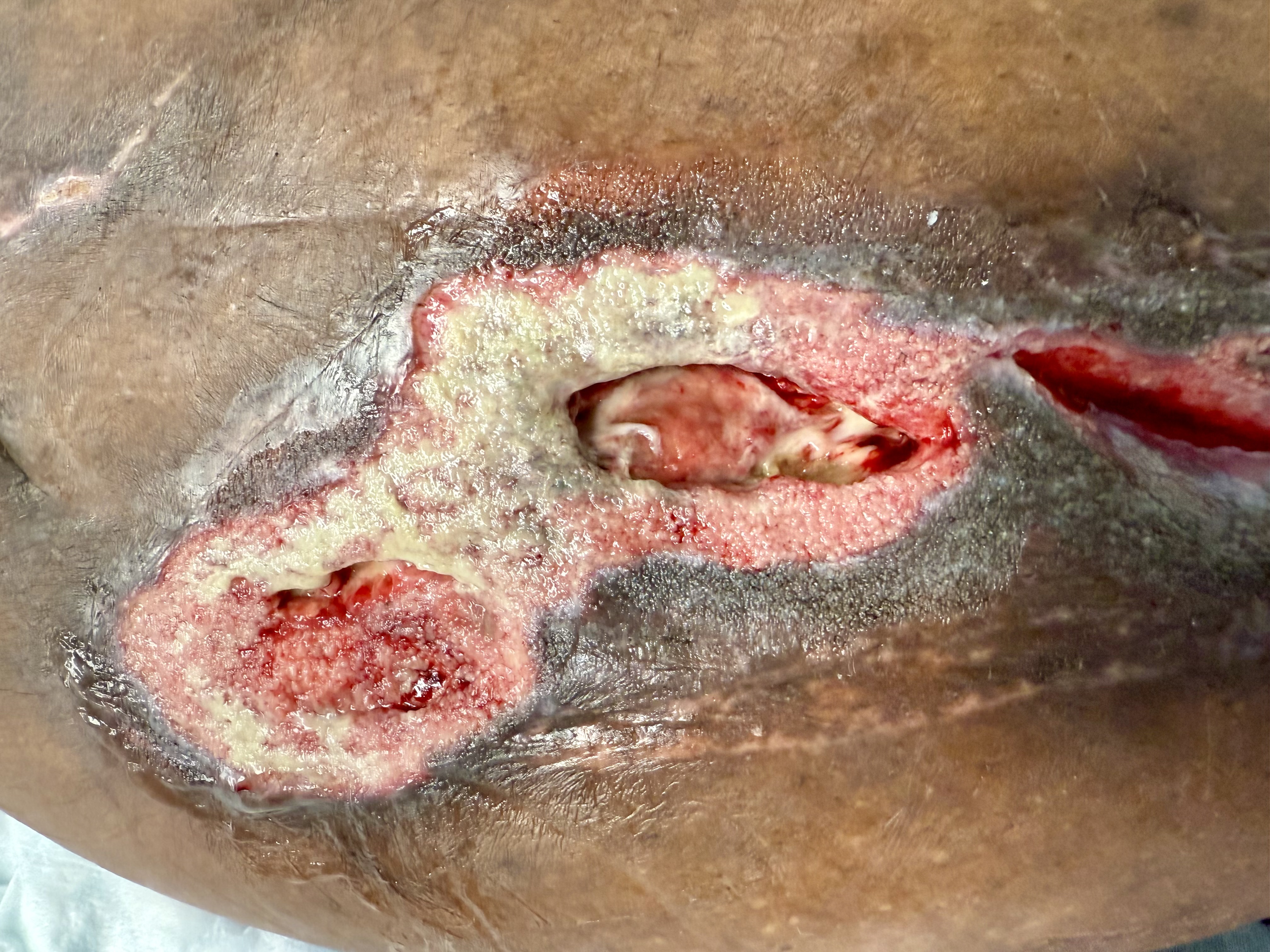
The patient was first evaluated at our wound center in December 2024. At that time, painful pannicular ulcers were present at prior biopsy sites, demonstrating full-thickness skin loss with extension into the subcutaneous tissue (Figure 1). No surrounding cellulitis, sinus tracts, or tunneling were identified on examination. There were no areas of expressible purulence.
FIGURE 1 Left lower quadrant wounds developed after painful subcutaneous nodules.
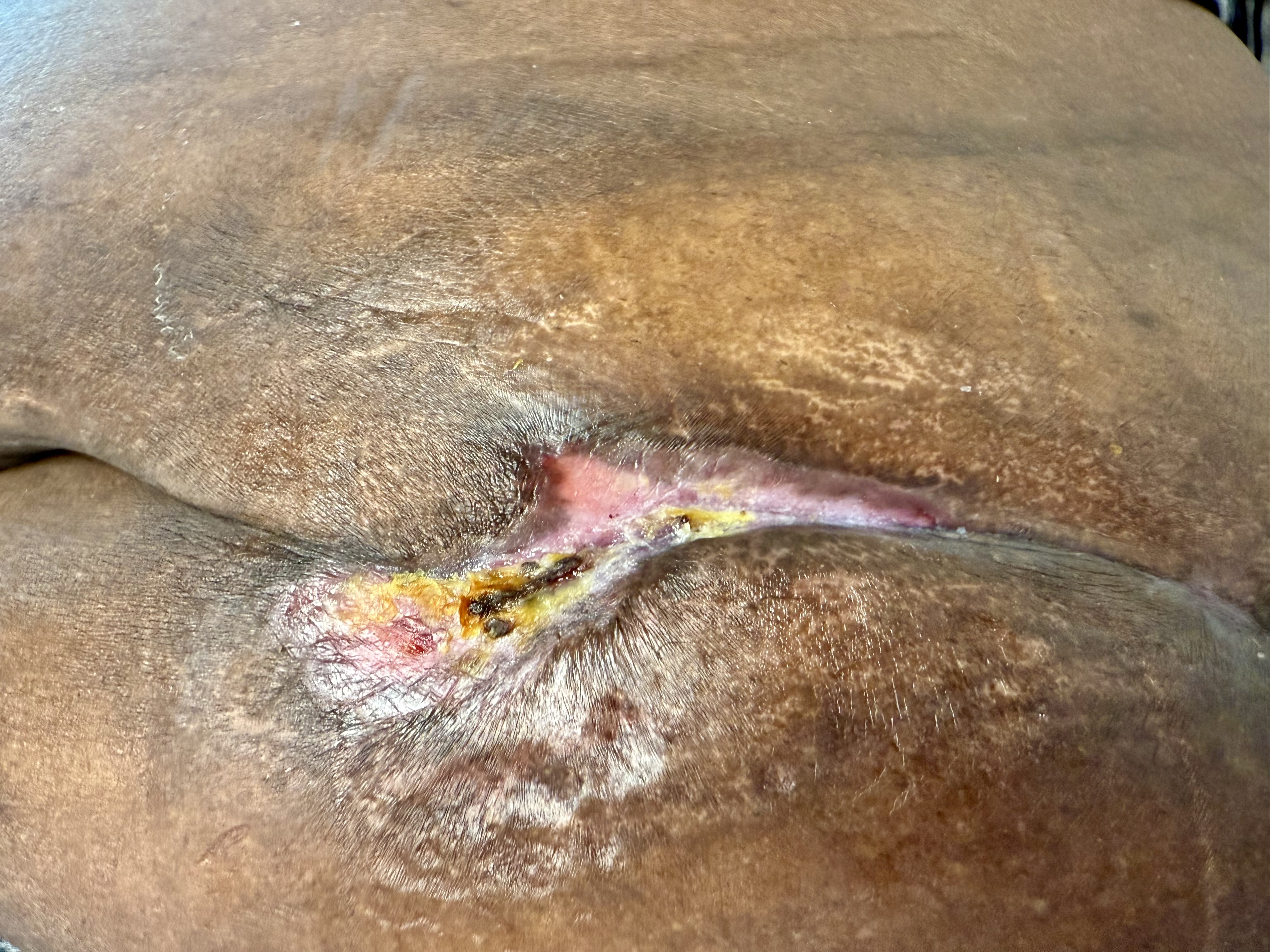
Gradual wound improvement was observed through early 2025, and by April 2025, near-complete epithelialization had been achieved (Figure 2). Shortly thereafter, complete closure was achieved. STS therapy was discontinued by Nephrology.
FIGURE 2 Complete closure within 2 weeks of this image.
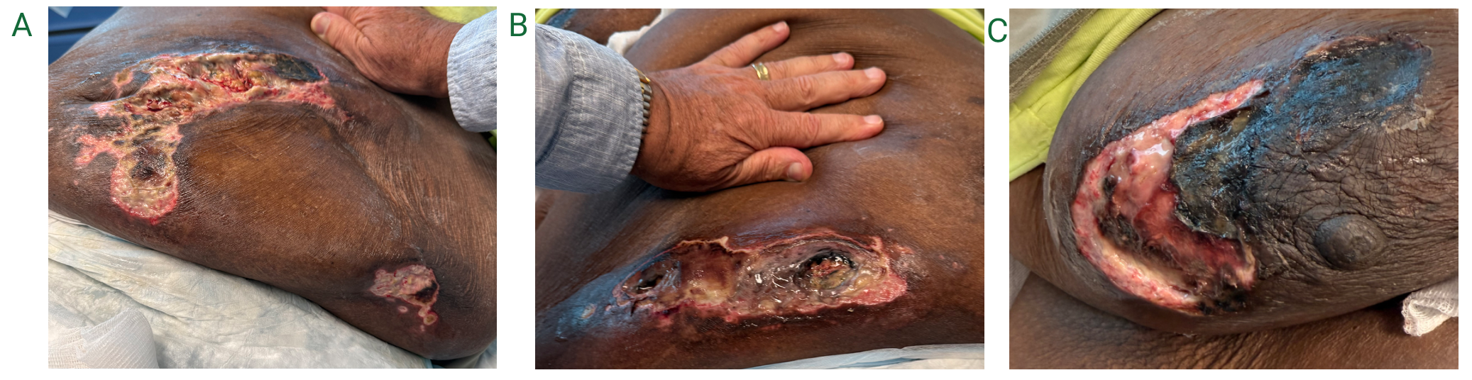
In June 2025, recurrence occurred in the left lower abdominal pannus (Figure 3). Despite continued wound care, the wound progressed. By mid-September 2025, extensive bilateral pannicular necrosis developed with involvement of the right breast (Figure 4A–C). Debridement revealed widespread liquefactive adipose necrosis beneath devitalized skin. Spreading lesions were again characterized by dry black skin that when unroofed exposed larger areas in the adipose layer below and extending outward with liquefactive necrosis. Debridement in the outpatient setting was not well tolerated and was therefore deferred while inpatient admission was arranged.
FIGURE 3 Recurrence after stopping sodium thiosulfate.
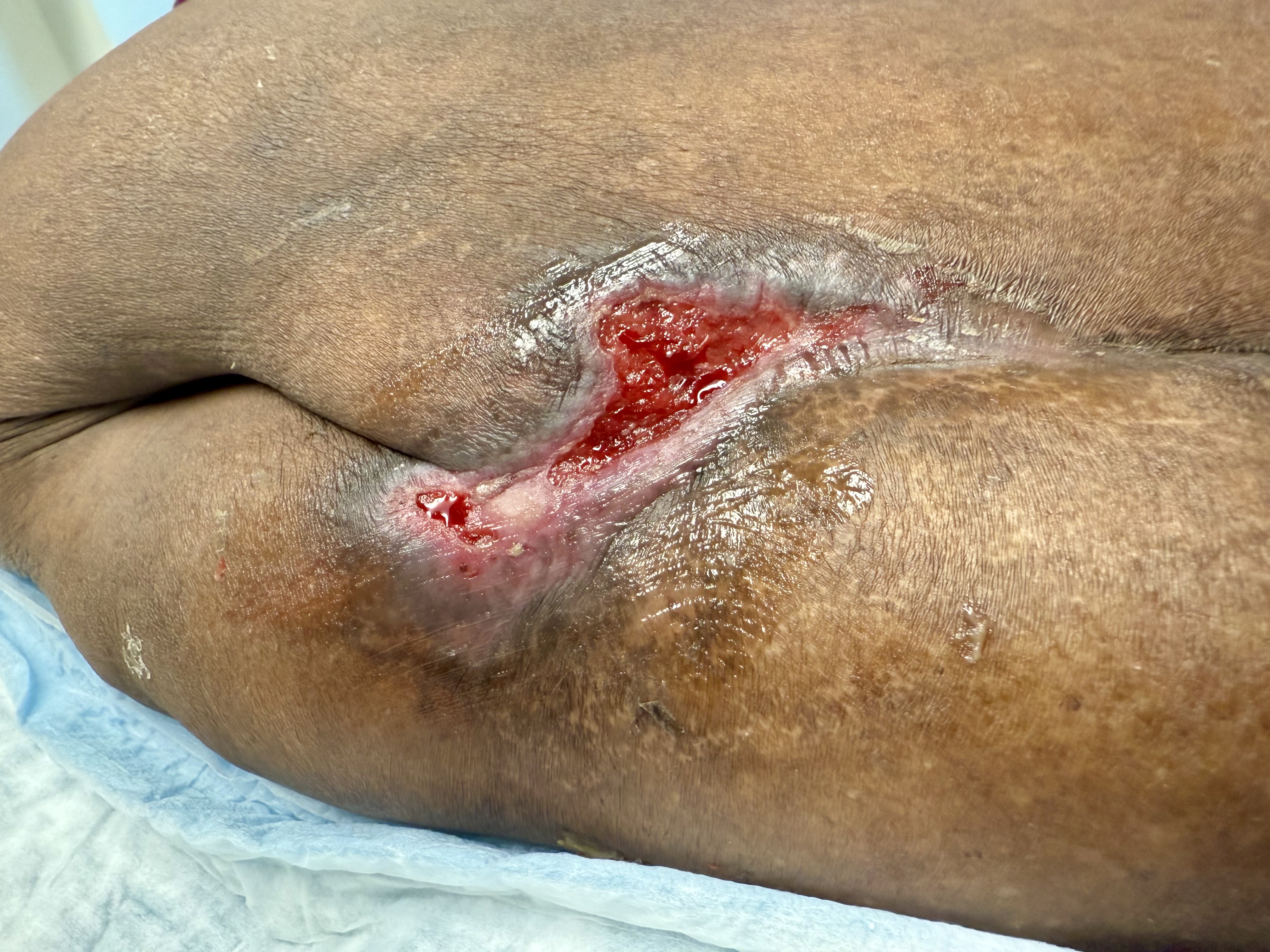
FIGURE 4 A-C) Extensive right and left lower quadrant pannicular breakdown to include right breast as well.
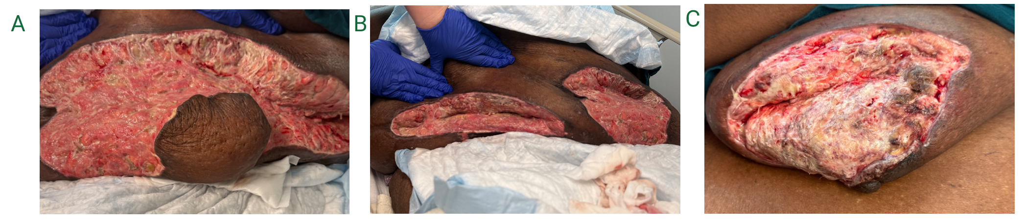
The patient underwent deep and wide operative debridement with removal of necrotic skin and adipose tissue (Figure 5A-C). Negative pressure wound therapy (NPWT) was initiated postoperatively. Due to wound complexity, she required approximately 60 days of skilled nursing facility management. Selected antimicrobial dressings were utilized during the final epithelialization phase. By January 14, 2026, complete healing was observed without evidence of recurrent necrosis.
FIGURE 5 After extensive wide and deep operative debridement for source control. A) Left lower quadrant. B) Right lower quadrant with left lower quadrant visible at left edge. C) Right breast.
Results
Application of systemic therapy, serial debridement, operative source control, NPWT, and subsequent antimicrobial dressing selection resulted in progressive wound stabilization and eventual closure. Following the initial course of STS therapy and structured wound management, nearly complete epithelialization was achieved by April 2025. Shortly thereafter, complete closure was achieved. After recurrence in June 2025 and subsequent progression to extensive pannicular and breast involvement, aggressive surgical debridement with removal of necrotic skin and liquefactive adipose tissue was required.
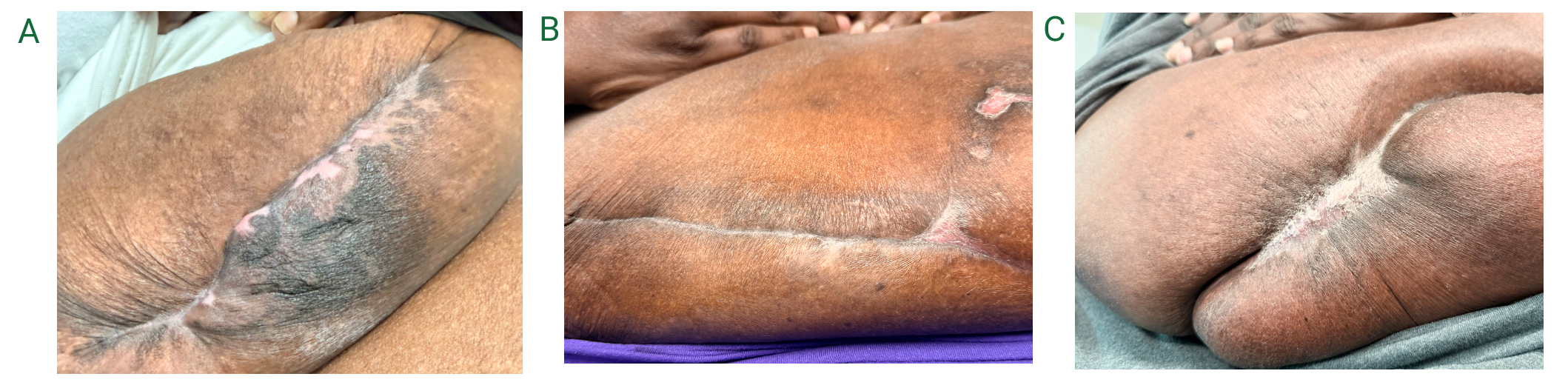
Postoperatively, NPWT facilitated granulation tissue formation and reduction in wound volume. Transition to advanced antimicrobial dressings during the remodeling phase supported continued epithelial advancement. By January 14, 2026, all wounds demonstrated intact epithelialization with no wounds demonstrating evidence of ongoing necrosis, active infection, or further recurrence (Figure 6A-C).
FIGURE 6 A-C) All three areas with complete closure.
At final follow-up, the patient remained clinically stable, without additional pannicular lesions or signs of progressive calciphylaxis.
Discussion
This case demonstrates classic features of calciphylaxis, including severe pain preceding visible breakdown, adipose-predominant involvement, progressive ischemic necrosis, and liquefactive degeneration of subcutaneous fat. Obesity likely contributed to both the distribution and severity of the disease because of impaired microvascular perfusion and increased oxygen diffusion distance within adipose-rich areas.7
The absence of consistent cellulitis or tunneling suggests that microvascular thrombosis, rather than infection, was the primary driver of tissue destruction. Warfarin-associated skin necrosis was considered less likely because the patient was not receiving warfarin. Vasculitis also was considered less likely because petechiae, purpura, and other characteristic vasculitic features were not observed clinically. An abdominal plain film obtained during hospitalization noted vascular calcifications; however, this finding was considered supportive rather than diagnostic in isolation.
Pathology findings from the November 2025 extensive debridement did include the following: “Ulcer and gangrenous necrosis, focal calcification in vascular walls”. Comment further noted, “intermediate sized vessels…this may suggest calciphylaxis; clinical correlation is recommended.” There was a clear temporal relationship between clinical improvement and STS initiation as well as recurrence with abrupt discontinuation. Once she completed healing the recurrent wounds, she was not taken off STS, rather she was begun on a very slow taper. Although STS therapy appeared temporally associated with stabilization, definitive control required aggressive surgical source control.
A structured wound management framework incorporating systemic optimization, infection surveillance, serial debridement, NPWT, and dressing selection based on wound characteristics facilitated progressive granulation and closure. This approach ensured systematic attention to key barriers to healing while underscoring the importance of multidisciplinary coordination among nephrology, surgery, infectious disease, and wound specialists. Even severe recurrent calciphylaxis may achieve complete healing when both systemic and local drivers are addressed.
Conclusion
Calciphylaxis remains a diagnostically complex and therapeutically challenging disorder associated with high morbidity. This case highlights the pathogenic contribution of obesity in pannicular disease, in particular, the limitations of biopsy confirmation (not seen in the punch biopsies but later described in the larger tissue specimen), and the necessity of aggressive surgical intervention in advanced presentations. Even in severe recurrent disease, sustained multidisciplinary management within a structured wound care framework may result in meaningful wound resolution.