Introduction
Chronic wounds represent a significant clinical challenge, particularly among patients with multiple comorbidities that impair normal tissue repair. One of the most prevalent and debilitating chronic wound types is the pressure ulcer (PrU) (also known as a pressure injury), which develops when sustained mechanical forces compromise perfusion and tissue integrity at bony prominences such as the sacrum, heels, and ischial tuberosities.1 The National Pressure Injury Advisory Panel (NPIAP) defines pressure ulcers as localized injuries affecting the skin and underlying tissues.2 Limited mobility is a major risk factor for pressure ulcers, and its presence often complicates both prevention and treatment strategies. The risk of developing a PrU is influenced by various factors, including the duration of pressure, the intensity of pressure, and the individual’s overall health status, making it a significant concern across diverse healthcare settings, including acute care hospitals, long-term care and skilled nursing facilities, inpatient rehabilitation centers, home health services, hospice and palliative care programs, and outpatient wound care clinics.3
Standard treatment protocols for PrUs emphasize pressure offloading/pressure redistribution, moisture balance, infection control, nutritional optimization and debridement to prepare the wound bed for healing.4 While these measures are foundational, they often fail to achieve closure in recalcitrant wounds, particularly in individuals with complex comorbid profiles.5 Moreover, despite the availability of clinical guidelines and preventive strategies, current research indicates that the global incidence of PrUs is on the rise.6
Between 1990 and 2021, the global incidence of PrUs more than doubled, rising from approximately 1.14 million to 2.47 million cases.1 Although age-standardized incidence rates (ASIR) have remained relatively stable, the absolute burden reflected in mortality and disability-adjusted life years (DALYs) has increased significantly.1
While effective management requires timely intervention, patients with restricted mobility frequently encounter barriers to accessing care, including transportation difficulties, financial constraints, and lack of supportive resources. Thus, PrUs have remained a persistent global health challenge, particularly among aging populations and individuals with chronic conditions such as diabetes, cardiovascular disease, and neurological impairments. Recent findings from the Global Burden of Disease Study 2021 underscore the escalating impact of PrUs on public health systems worldwide.1 Cellular, acellular, and matrix-like products (CAMPs), such as placental-derived allografts, have emerged as promising adjuncts to conventional care. These grafts provide a biologically active scaffold enriched with growth factors and cytokines that support angiogenesis, modulate inflammation, and promote re-epithelialization.7 Evidence from studies on diabetic foot ulcers and venous leg ulcers suggests that placental-based products can accelerate healing and reduce complications; however, their role in managing pressure ulcers remains underexplored.8
A retrospective pilot feasibility study was conducted to evaluate the use of an amniotic tissue membrane (ATM) for the treatment of PrUs using real-world evidence. Specifically, the authors assessed the effectiveness of Membrane Wrap™ (BioLab Holdings, Mesa, Arizona) within routine wound care practice.
This study, which is based on retrospectively gathered data from multiple healthcare sites, offers valuable real-world insights regarding the clinical effectiveness of these products in managing chronic PrUs in a complex and geographically varied patient population.
Methods
Study design and oversight
This multicenter, retrospective cohort study evaluated the effectiveness of a human ATM compared with standard of care (SOC) in the treatment of PrUs. SOC consisted of clinician-directed PrU management documented within the medical record and typically included pressure redistribution/offloading, support surfaces, wound cleansing, debridement when indicated, moisture-balancing dressings, infection management, nutritional support, and other therapies deemed appropriate by the treating clinician. Because treatment was evaluated in a real-world retrospective dataset, the precise combination and intensity of SOC interventions varied among patients and sites.
The study was conducted using de-identified real-world electronic health record (EHR) data under a waiver of informed consent and HIPAA authorization granted by the Sterling Institutional Review Board (IRB #13631; April 23, 2025). All data handling procedures adhered to applicable ethical and data protection standards.
Data sources
Data were extracted from the Intellicure EHR system for the period February 2017 through December 2025. Clinical variables were obtained from the U.S. Wound Registry (USWR), a national repository of research grade wound care data (Intellicure, LLC; The Woodlands, TX). Patients received either ATM plus SOC or SOC alone. The USWR accumulates clinical data through a purpose-built EHR system (Intellicure, LLC) utilized by over 1000 health care practitioners across 29 states.
Data quality and case selection
Data quality was ensured through manual validation performed by trained abstractors. Outliers and missing values were retained to preserve the robustness and representativeness of the dataset. Therefore, patients were not excluded even if wound treatment outcome data were unavailable due to death, transfer of care, or loss to follow-up.
For the treatment arm (ATM plus SOC), each patient contributed only a single Index Wound, defined as the largest wound by surface area at the time of the first ATM application. For the control arm (SOC only), the Index Wound was defined as the largest wound by surface area at the time of the first wound size measure. Wounds were excluded if they (1) had misleading or incorrect location descriptors (e.g., “breast,” “vaginal”), (2) were ≤1 cm2 in size, (3) might not be appropriate for CAMP treatment (i.e., tissue type exposed labelled as “intact skin” or “partial thickness”), or (4) did not have the CAMP of interest as the final cellular or tissue based product applied.
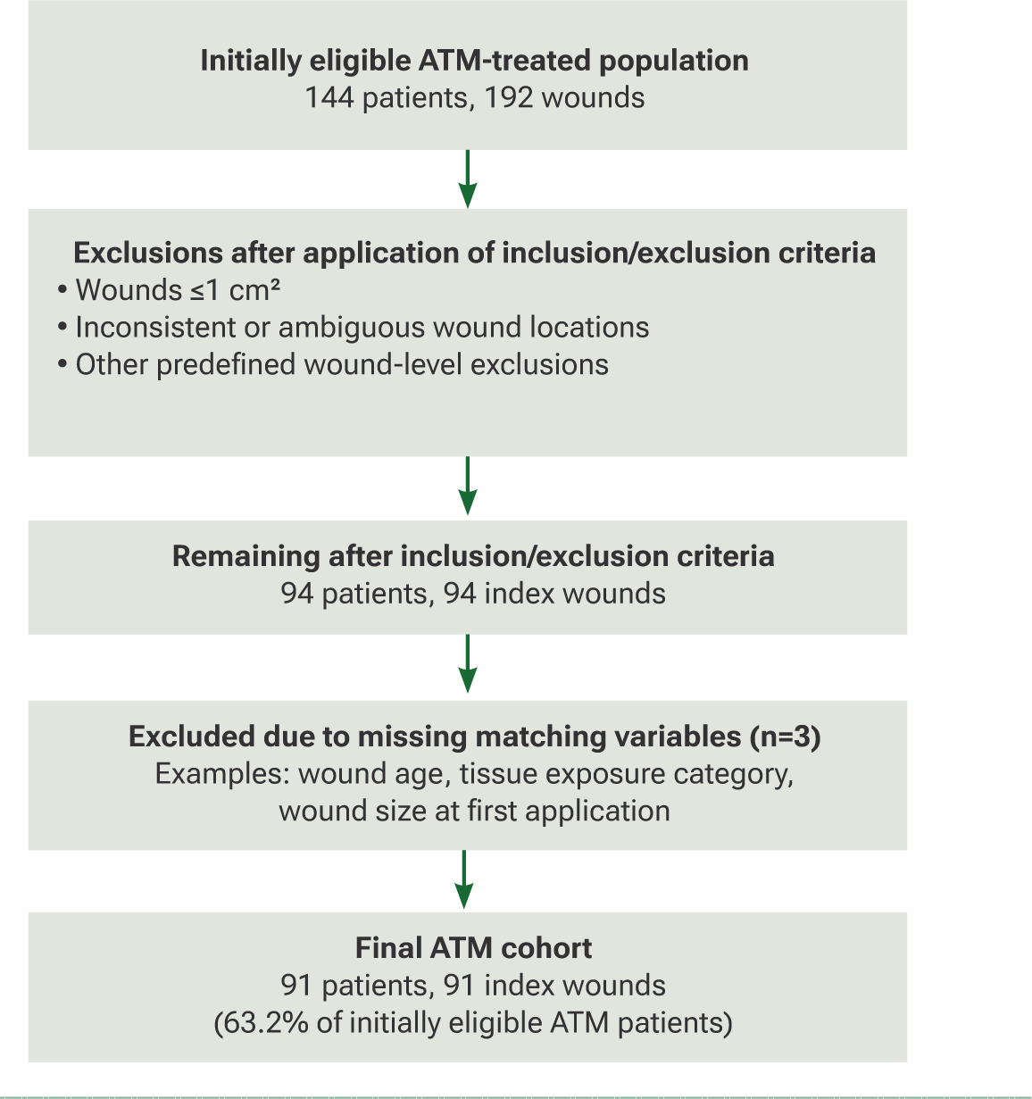
Cohort derivation is summarized in Figure 1. A total of 144 ATM-treated patients contributing 192 wounds were initially identified as eligible for review. Following application of study-specific inclusion and exclusion criteria, including removal of wounds ≤1 cm2 and wounds with inconsistent or ambiguous location descriptions, 94 patients with 94 index wounds remained. An additional three patients were excluded because of missing values in variables required for matching, resulting in a final ATM cohort of 91 patients with 91 index wounds. No additional ATM patients were excluded during the matching process.
FIGURE 1 Study cohort derivation. Flow diagram showing identification of eligible ATM-treated patients and wounds, application of study inclusion/exclusion criteria, exclusion due to missing matching variables, and formation of the final analytical ATM cohort (91 patients, 91 index wounds; 63.2% of initially eligible ATM patients).
Exposure groups
Subjects were classified into two cohorts:
-
ATM group: received human amniotic tissue membrane in addition to SOC.
-
SOC group: received SOC alone without any CAMPs.
Data processing and variable definitions
Patient-level variables
Patient age was recalculated relative to the date of the first visit for the Index Wound. The total number of concomitant wounds (one of the matching factors that reflects condition severity) was re-categorized into: no concomitant wounds (0), a few concomitant wounds (1-3), and many concomitant wounds (4 or more). Comorbidities included diabetes, chronic kidney disease, congestive heart failure, peripheral artery disease, autoimmune disease, dementia, and paralysis.
Wound-level variables
Wound location was determined using ICD-10 codes L89.0–L89.9. When multiple tissues were exposed, the more severe exposure category was selected (e.g., bone superseded tendon). Wound stage was redefined based on tissue exposure:
-
Stage 4: bone, tendon, muscle, fascia, or other deep tissues
-
Stage 3: joint capsule, adipose, subcutaneous tissue
-
Stage 2: partial thickness wounds
-
Stage 1: intact skin or indeterminate stage.
Outcome definitions and imputation
Wound outcomes with a descriptor “healed” or “resolved” were deemed as Healed. For uncertain wound outcome descriptors (e.g., “improving”, “worse”, “transferred”, and “lost to follow-up”), percentage area reduction (PAR) was used to assign outcome categories using prespecified rules:
-
Wounds achieving ≥97% area reduction at last measurement were imputed as Healed.
-
Based on the average PAR over a 100 day evaluation window (reflecting the empirically observed mean episode duration), outcomes were imputed as:
-
No change: in between ±10% change
-
Healing: >10% reduction
-
Worse: >10% increase in wound size
-
Patients who transferred care, were lost to follow-up, died, or otherwise lacked a definitive healing status were retained in the dataset to preserve the real-world nature of the cohort and reduce survivorship bias. When sufficient wound measurement data were available, prespecified PAR criteria were used to assign wound trajectory categories. Cases lacking adequate information for outcome determination were retained and reported as missing outcomes.
Because definitive healing status was not consistently available in the electronic health record, prespecified operational definitions were used to categorize wound trajectories. A PAR ≥97% was classified as healed to account for minor residual wound measurements and variability in routine clinical documentation and measurement variability. Average PAR over the evaluation period was further used to categorize wounds as healing (>10% reduction), no change (within ±10%), or worse (>10% increase). These thresholds were developed as pragmatic study-specific rules to support analysis of real-world data and should not be interpreted as validated clinical definitions of healing or treatment response.
Matching procedure
A matching model was employed to reduce confounding between treatment groups. Factors used in the matching process included patient age, sex, arrival method, wound location, tissue exposure type, wound age at first CAMP application, wound size at first CAMP application, number of concomitant wounds, and all recorded comorbidities. Mahalanobis distance-based nearest neighbor matching was applied using R version 4.5.1 and the MatchIt package version 4.7.2.
Statistical analysis
Descriptive statistics summarized baseline characteristics of the matched cohorts. For matched datasets, paired analyses were performed:
-
Continuous variables: Wilcoxon signed rank test
-
Categorical variables: McNemar test All tests were two sided with a Type I error rate of 0.05. Analyses were conducted in R software for matched observational study design.
Results
Baseline demographic and clinical characteristics
Patient selection is illustrated in Figure 1. Of the 144 initially eligible ATM-treated patients, 91 (63.2%) remained in the final matched cohort after application of study inclusion/exclusion criteria and removal of cases with missing matching variables. The matching procedure itself did not result in further loss of ATM patients. The groups were well balanced across most demographic and clinical covariates (Table 1).
TABLE 1 Prevalence of comorbidities by treatment group
| Comorbidity | ATM (%) | SOC (%) | P-value |
|---|---|---|---|
| Diabetes | 33.0 | 33.0 | 1.0000 |
| Chronic kidney disease | 15.4 | 14.3 | 1.0000 |
| Congestive heart failure | 18.7 | 8.7 | — |
| Dementia | 19.8 | 17.6 | 0.4795 |
| Peripheral artery disease | 13.2 | 11.0 | 0.4795 |
| Tobacco use | 7.7 | 7.7 | — |
| Obesity | 9.9 | 15.4 | 0.3320 |
| Depression | 9.9 | 12.1 | 0.8137 |
| Atrial fibrillation | 9.9 | 12.1 | 0.7893 |
| Osteomyelitis | 4.4 | 3.3 | 1.0000 |
| Hyperlipidemia | 12.1 | 16.5 | 0.5403 |
| Hypertension | 38.5 | 47.3 | 0.3123 |
| Paralysis | 18.7 | 15.4 | 0.2482 |
ATM, amniotic tissue membrane; SOC, Standard of care. “—” indicates p-values not reported (identical prevalence between groups). Values are presented as percentages
Age and sex
Mean age was similar between groups (ATM 74.15 ± 14.20 years vs SOC 74.76 ± 12.63 years; p=0.3276). The proportion aged ≥65 years did not differ (ATM 81.3% vs SOC 80.2%; p=1.0000). Sex distribution was comparable (female: ATM 48.4% vs SOC 46.2%; p=0.7237).
Race and BMI
Recorded race categories appeared similar among those with available data; however, race was frequently missing (ATM n=76; SOC n=59), limiting interpretability of between group comparisons. BMI was also missing for many participants (ATM n=75; SOC n=20). Among those with data, BMI trended higher in the ATM group (mean ± SD: 27.62 ± 8.71 vs 24.89 ± 6.33; p=0.4144), but this difference did not reach statistical significance.
Functional status (arrival method) and concomitant wounds
Arrival method categories (ambulatory, wheelchair, stretcher/bedridden) were closely matched with no evident differences across groups, and no missing data were reported for this variable. The distribution of concomitant wound counts (0, 1-3, or 4+) was also similar between cohorts.
Comorbidities
The prevalence of key comorbidities was broadly comparable: diabetes (ATM 33.0% vs SOC 33.0%; p=1.0000), chronic kidney disease (ATM 15.4% vs SOC 14.3%; p=1.0000), congestive heart failure (both 18.7%), dementia (ATM 19.8% vs SOC 17.6%; p=0.4795), peripheral artery disease (ATM 13.2% vs SOC 11.0%; p=0.4795), tobacco use (both 7.7%), obesity (ATM 9.9% vs SOC 15.4%; p=0.3320), depression (ATM 9.9% vs SOC 12.1%; p=0.8137), atrial fibrillation (ATM 9.9% vs SOC 12.1%; p=0.7893), osteomyelitis (ATM 4.4% vs SOC 3.3%; p=1.0000), hyperlipidemia (ATM 12.1% vs SOC 16.5%; p=0.5403), and hypertension (ATM 38.5% vs SOC 47.3%; p = 0.3123), and paralysis (ATM 18.7% vs SOC 15.4%; p=0.2482) (Table 1). No missingness was reported for comorbidity variables.
Overall, between the ATM and SOC groups, baseline demographic and clinical characteristics were well matched. Substantial missingness in race (especially in ATM) and BMI (particularly in ATM) should be considered when interpreting subgroup or adjusted analyses involving these variables.
Baseline wound characteristics
At baseline, wound characteristics were comparable between patients in the ATM group and in the SOC group (Table 2). Pressure ulcer staging was distributed similarly across groups, with Stage 3 ulcers comprising 53.8% of wounds in the ATM group and 54.9% in the SOC group, and Stage 4 ulcers comprising 46.2% and 45.1%, respectively. Wound location patterns were also consistent between treatment arms. The most common site of ulceration was the back (40.7% in both groups), followed by the heel and midfoot (20.9% in ATM; 19.8% in SOC) and the buttock (15.4% in ATM; 16.5% in SOC). Less frequent locations included the hip (9.9% in both groups), ankle (3.3% in each group), contiguous back/buttock/hip sites (1.1% in both groups), other uncovered sites (7.7% in both groups), and sites listed as unspecified (1.1% in both groups).
TABLE 2 Wound characteristics of pressure ulcers
| Characteristic | No. (%) of patients in ATM group (n=91) | No. (%) of patients in SOC group (n=91) | P-value |
|---|---|---|---|
| Ulcer stage | 1.0000 | ||
| Stage | 3 49 (53.8%) | 50 (54.9%) | |
| Stage | 4 42 (46.2%) | 41 (45.1%) | |
| Wound location NaN | |||
| Ankle | 3 (3.3%) | 3 (3.3%) | |
| Back | 37 (40.7%) | 37 (40.7%) | |
| Buttock | 14 (15.4%) | 15 (16.5%) | |
| Contiguous site of back, buttock, and hip | 1 (1.1%) | 1 (1.1%) | |
| Heel and midfoot | 19 (20.9%) | 18 (19.8%) | |
| Hip | 9 (9.9%) | 9 (9.9%) | |
| Other site uncovered | 7 (7.7%) | 7 (7.7%) | |
| Unspecified site | 1 (1.1%) | 1 (1.1%) | |
| Worst tissue type exposed NaN | |||
| Bone | 8 (8.8%) | 9 (9.9%) | |
| Muscle | 26 (28.6%) | 24 (26.4%) | |
| Subcutaneous | 49 (53.8%) | 50 (54.9%) | |
| Tendon | 8 (8.8%) | 8 (8.8%) | |
| Wound depth >1 cm | 27 (30.0%) | 23 (25.8%) | .5708 |
| Unknown | 1 (1.1%) | 2 (2.2%) | |
| Granulation tissue percentage NaN | |||
| 0% | 0 (0.0%) | 5 (6.9%) | |
| >0% and <25% | 13 (15.9%) | 11 (15.3%) | |
| Between 25% and 50% | 14 (17.1%) | 8 (11.1%) | |
| >50% and <100% | 35 (42.7%) | 27 (37.5%) | |
| 100% | 20 (24.4%) | 21 (29.2%) | |
| Unknown | 9 (9.9%) | 19 (20.9%) | |
| Wound on the lower extremity (leg, foot, or heel) | .3506 | ||
| Mean (SD) | 0.4 (0.5) | 0.3 (0.5) | |
| Median (Q1, Q3) | 0.0 (0.0, 1.0) | 0.0 (0.00, 1.0) | |
| Wound time in service from 1st visit to 1st application (days) | .0111 | ||
| Mean (SD) | 26.9 (92.5) | 41.3 (108.6) | |
| Median (Q1, Q3) | 0.0 (0.0, 20.0) | 14.0 (0.0, 35.0) | |
| Wound age at 1st application (days) | < 1e-04 | ||
| Mean (SD) | 329.5 (974.2) | 220.6 (853.4) | |
| Median (Q1, Q3) | 105.0 (65.0, 326.0) | 69.00 (28.5, 133.5) | |
| Wound age at 1st application (cm2) | .0275 | ||
| Mean (SD) | 18.9 (31.4) | 14.2(21.3) | |
| Median (Q1, Q3) | 8.1 (5.0, 22.9) | 7.2 (2.4, 16.6) | |
| No. of concomitant wounds per patient | .NaN | ||
| 0 | 48 (52.7%) | 47 (51.6%) | |
| 1-3 | 40 (44.0%) | 41 (45.1%) | |
| 4+ | 3 (3.3%) | 3 (3.3%) |
The deepest tissue type exposed within each ulcer did not differ meaningfully between groups. Subcutaneous tissue was the most frequently exposed structure (53.8% ATM; 54.9% SOC), followed by muscle (28.6% ATM; 26.4% SOC). Bone exposure was observed in 8.8% of ATM patients and 9.9% of SOC patients, while tendon exposure occurred in 8.8% of patients in each group. Wound depth >1 cm was present in 30.0% of ATM and 25.8% of SOC wounds. Granulation tissue percentage at baseline varied widely across patients but demonstrated similar distributions between groups. In the ATM and SOC groups, respectively, 0% granulation was present in 0% of ATM subjects vs. 6.9% of wounds in the SOC arm; >0–25% in 15.9% vs. 15.3%; 25–50% in 17.1% vs. 11.1%; >50–100% in 42.7% vs. 37.5%; and 100% granulation in 24.4% vs. 29.2%. Missing estimates were more frequent in the SOC group (20.9%) compared with the ATM group (9.9%).
Lower-extremity wound involvement (leg, foot, or heel) was low overall and similar between arms, with mean (SD) values of 0.4 (0.5) wounds in the ATM group and 0.3 (0.5) in the SOC group.
Measures of wound chronicity, defined as the duration of the wound prior to initiation of documented wound therapy in the medical record, showed some baseline differences between groups. The time from first clinic visit to first application was shorter among ATM patients (mean 26.9 days) than SOC patients (mean 41.3 days). In contrast, wound duration prior to treatment initiation was longer in the ATM group (median 105 days) compared with the SOC group (median 69 days), although both groups demonstrated substantial variability. Median values were reported for wound duration due to a skewed distribution.
Wound size at first application also differed modestly, with ATM patients exhibiting a larger mean wound area (18.9 cm2) than SOC patients (14.2 cm2). Concomitant wound burden was similar between groups: approximately half of patients in each group had no additional wounds (52.7% ATM; 51.6% SOC), while 44.0% of ATM and 45.1% of SOC patients had one to three additional wounds, and 3.3% in each group had four or more.
Changes in clinical characteristics of the wounds
Across both treatment groups, overall trends show modest changes in wound size but marked differences in practitioner-assessed outcomes. Notably, the ATM group demonstrates a higher proportion of wounds rated as improved or healing, whereas the SOC group shows more cases with unknown, transferred, or lost-to-follow-up outcomes (Table 3).
TABLE 3 Wound outcomes of pressure ulcers.
| Characteristic | No. (%) of patients in ATM group (n=91) | No. (%) of patients in SOC group (n=91) | P-value |
|---|---|---|---|
| Percent Wound Size Change | .7994 | ||
| Mean (SD) | -0.1 (1.1) | 0.1 (2.4) | |
| Median (Q1, Q3) | -0.3 (-0.8, 0.0) | -0.2 (-0.7, 0.0) | |
| Unknown | 0 (0.0%) | 3 (3.3%) | |
| Wound Outcome (by Practitioner) | NaN | ||
| Better | 46 (52.3%) | 18 (25.7%) | |
| Death | 0 (0.0%) | 5 (7.1%) | |
| Healed | 12 (13.6%) | 26 (37.1%) | |
| Lost to Follow-Up | 0 (0.0%) | 6 (8.6%) | |
| No Change | 5 (5.7%) | 1 (1.4%) | |
| Transferred Care | 2 (2.3%) | 7 (10.0%) | |
| Worse | 23 (26.1%) | 7 (10.0%) | |
| Unknown Outcome | 3 (3.3%) | 21 (23.1) | |
| Wound Time in Service since 1st Application (days) | .2828 | ||
| Mean (SD) | 67.0 (66.5) | 100.7 (200.1) | |
| Median (Q1, Q3) | 56.0 (28.0, 76.5) | 23.0 (2.0, 86.0) | |
| Wound Outcome Based on PAR | .0449 | ||
| Healing or Healed | 58 (63.7%) | 44 (48.4%) | |
| No Change, Worse, or Not Healed | 33 (36.3%) | 47 (51.6%) |
ATM, human amniotic tissue membrane + standard of care; NaN, P value not available due to numeric issues (not enough sample); PAR, percentage area reduction; Q1, 1st quartile 1; Q3, 3rd quartile; SD, standard deviation; SOC, standard of care alone
Granulation tissue and depth
The distribution of granulation coverage trended toward more intermediate coverage (50–100%) in the ATM arm (42.7% vs 37.5%), whereas the SOC arm showed a larger fraction at 100% and at 0%; however, the difference did not reach statistical significance. Interpretation is limited by missing data (ATM n=9; SOC n=19). Wounds deeper than 1 cm were similar between groups (ATM 30.0% vs SOC 25.8%; p=0.5708).
Time in care and timing of first application
From the first clinic visit to the first CAMP application, ATM wounds had substantially shorter time in service (mean ± SD 26.86 ± 92.49 days; median 0 [0, 20]) than SOC (41.32 ± 108.62 days; median 14 [0, 35]; p=0.0111), indicating earlier intervention in the ATM arm. Conversely, the wound age at first application was older in ATM (median 105 days [65, 326]) versus SOC (69 days [29, 134]; p<1×10⁻⁴), suggesting that although ATM was applied earlier after presentation, these wounds had been present longer prior to index care. After first application, time in service until outcome assessment was comparable between groups (ATM 66.97 ± 66.53 days vs SOC 100.73 ± 200.07 days; p=0.2828).
Wound size dynamics
Percent change in wound size over follow-up did not differ statistically between groups (mean ± SD: ATM −0.08 ± 1.15 vs SOC 0.06 ± 2.41; p=0.7994), with similar median changes (ATM −0.27 [−0.78, 0.00] vs SOC −0.17 [−0.73, 0.00]). Despite the lack of statistical significance, the ATM group demonstrated a numerically greater reduction in wound size compared with SOC.
As shown in Figure 2, mean percent area reduction (PAR) trajectories with 95% confidence intervals were plotted for both groups. The ATM group had follow-up data available for a shorter duration (up to approximately 500 days), whereas the matched SOC group had data extending to approximately 900 days. This difference reflects variation in follow-up time and data availability rather than a predefined study limit; in particular, it may be due to earlier wound resolution, loss to follow-up, or fewer long-term observations in the ATM group.
FIGURE 2 Smoothed mean curves of percentage area reduction (PAR) with 95% confidence intervals through 1,000 days. ATM, Human Amniotic Tissue Membrane + standard of care group; SOC, standard of care alone group.
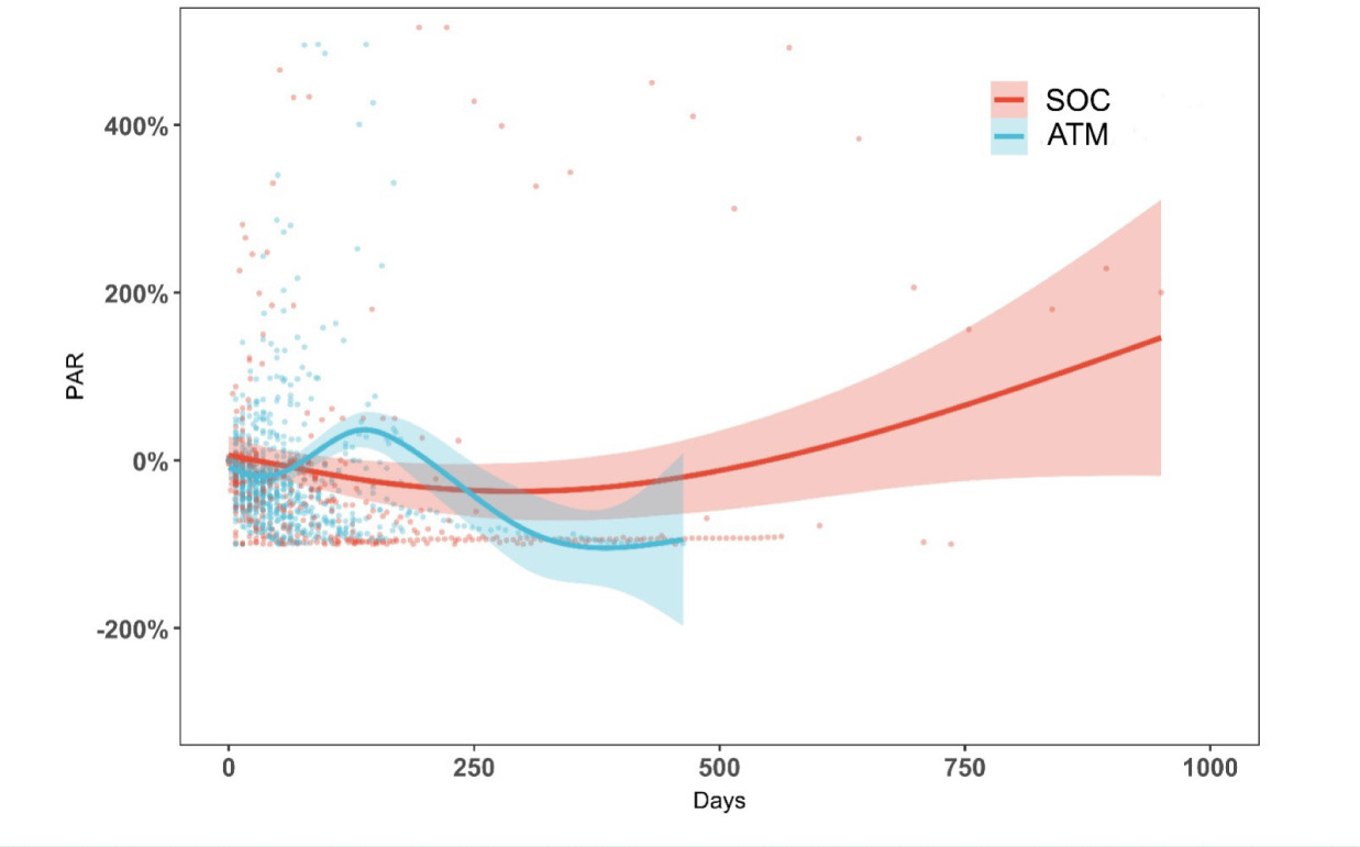
At Day 0 (i.e., the day of the first CAMP application), all the trajectories started at 0%, and the mean PAR trajectory of the SOC group decreased and then increased and ended at a positive PAR value, mainly due to the existence of a number of large wound size increases at different time points. The trajectory of the ATM group oscillated first and finally ended at a negative PAR value, suggesting a trend towards wound healing on average.
Composite clinical status at end of observation
When outcomes were grouped by clinical trajectory, the overall distribution differed significantly (p=0.0449): “Healing or Healed” occurred in 63.7% of ATM vs 48.4% of SOC and “No Change, Worse, Not Healed” in 36.3% of ATM vs 51.6% of SOC. There was a greater missingness in the SOC group in the detailed outcome description categories (SOC missing = 21 vs ATM missing = 3) and should be considered when interpreting comparative healing signals.
Wound stage, location, and tissue exposure
Stage distribution (Stage 2/3/4), anatomic location, and most severe tissue exposure were not statistically significantly different across arms by design of the matched dataset, indicating balanced structural wound severity at baseline for these domains.
Summary interpretation
In this matched cohort, ATM was applied earlier after presentation to care but to wounds that were older and slightly larger at first application. Subsequent percentage size reduction was comparable between groups, and this is an objective measure whereas composite outcome categories favored a higher proportion classified as “Healing or Healed” in ATM which is a subjective measure, counterbalanced by substantially more missing outcomes in SOC as well as an imbalance driven by transfers, losses to follow-up, and deaths recorded in that arm.
Discussion
PrU prevention and treatment strategies have traditionally been informed by studies conducted in hospitals and long-term care settings, leaving significant knowledge gaps regarding the experiences of patients managed in outpatient wound centers, home health, or broader community environments.9 This gap is particularly consequential because many individuals with PrUs, often older adults with multimorbidity, impaired mobility, and chronic wound histories, receive most of their wound care outside institutional settings.10 The lack of real world evidence from these care environments limits the ability of clinicians and policymakers to develop accessible, effective treatment pathways that reflect the complexity of wounds managed in everyday practice.
In the present study, we leveraged a large, national repository of research grade EHR data to evaluate clinical outcomes in a real world population characterized by advanced age, high comorbidity burden, and substantial functional limitations. By examining matched cohorts of patients treated with a human ATM and those receiving SOC, we sought to understand whether biologic augmentation confers measurable benefits in wound healing dynamics beyond conventional management.
Our analyses demonstrate that, despite substantial similarities in wound severity at baseline including identical distributions of stage, anatomic location, and tissue exposure, the two groups differed in several clinically relevant ways. Wounds in the ATM cohort were older and slightly larger at the time of first application, yet these wounds received biologic therapy earlier relative to presentation. Such patterns suggest that clinicians may selectively apply ATM earlier in the course of specialist care for wounds perceived as chronic, complex, or slow to progress. This real world treatment behavior underscores the importance of adjusting for wound chronicity and patient characteristics when evaluating comparative effectiveness.
Changes in clinical wound characteristics over time further illuminate the differing trajectories between groups. While percent area reduction did not significantly differ between ATM and SOC, granulation patterns and clinical outcome classifications suggest important qualitative distinctions. The ATM cohort exhibited higher proportions of wounds classified as “Healing or Healed,” whereas the SOC cohort showed a disproportionately large number of wounds with missing outcomes as well as transfer, loss to follow-up, or death. This imbalance in outcome availability highlights a critical challenge in real-world evidence studies: differential follow-up that may bias estimates of treatment effect if not addressed appropriately.
Although ATM was not associated with a statistically significant improvement in the objective measure of PAR, ATM-treated wounds were more frequently classified as healing or healed at the end of follow-up. This apparent discrepancy suggests that favorable findings were driven by clinician-assessed and imputed outcome classifications rather than objective wound size reduction. Accordingly, the results should be interpreted cautiously and viewed as suggestive of a potential association.
The use of real-world data is especially valuable in PrU research, as wound trajectories frequently deviate from linear healing models and are influenced by fluctuating comorbidities, social determinants of health, and care environment constraints.11 EHR derived datasets provide the granularity needed to evaluate these nonlinear pathways and to capture clinically meaningful outcomes such as tissue exposure changes, granulation progression, and time dependent wound size trajectories. Moreover, our matched cohort design mitigates confounding by balancing patient-level and wound-level characteristics, allowing for a more valid comparison of ATM versus SOC in a population reflective of real-world wound care.
Overall, this study contributes important real-world evidence on the utility of amniotic tissue grafts in managing complex PrUs in community based practice. While biologic membranes are frequently reserved for recalcitrant wounds, our findings suggest that their use even in older, chronically ill individuals may support favorable healing trajectories. These insights have practical implications for clinicians considering advanced therapies as well as policymakers tasked with determining coverage criteria that reflect real world need rather than idealized clinical trial populations.
Future research should apply survival or trajectory modeling to capture time to healing and wound progression inflection points. Further attention to outcome completeness and social determinants of health will strengthen the evidence base and enhance the development of equitable, effective treatment strategies for pressure ulcers across all care environments.
Limitations
This study has several important limitations that should inform the interpretation of our findings. Although the use of a large national EHR-derived dataset provides access to real-world outcomes across diverse care environments, important limitations related to data completeness and variable standardization remain. Because these data were derived from real-world electronic medical records rather than standardized randomized controlled trial data collection, variability and missingness in key variables are present, including inconsistencies in wound location documentation, Fitzpatrick skin type reporting, and the completeness of peripheral arterial disease (PAD) screening for all PrUs.
The retrospective design introduces potential biases inherent to observational research. Treatment assignment was not randomized; thus, despite the use of a robust matching approach, residual confounding may persist if unmeasured clinical or social variables influenced decisions to apply the amniotic membrane or standard care.
An additional limitation relates to the outcome imputation strategy. The PAR thresholds used to classify wounds as healed, healing, unchanged, or worse were study-specific operational definitions and were not derived from validated PrU outcome criteria. Alternative thresholds could have resulted in different outcome classifications and potentially different estimates of treatment effect. Consequently, findings based on these imputed outcome categories should be interpreted as exploratory.
Several wound-level variables exhibited substantial missingness, most notably outcome descriptors and granulation measurements. Outcome missingness was markedly higher in the SOC arm (21 cases) than in the ATM arm (3 cases). Because this imbalance may reflect differential loss to follow-up, transfer of care, or mortality, the possibility of informative missingness cannot be excluded. Consequently, comparative outcome estimates may be biased despite matching and other analytic adjustments.
Imbalances in missing data can distort comparisons, especially when unknown outcome categories (transfer, loss to follow-up, death) are disproportionately represented in one treatment group. Similarly, BMI and race were missing for large portions of the patient cohort, limiting the ability to generalize findings to demographic subgroups.
The wound size data available for this analysis represent summary measures (e.g., size at first application, percent area change) rather than visit level time series measurements. As a result, the study could not evaluate healing trajectories, time to 50% reduction, or non-linear wound healing patterns, an important limitation given the complex trajectory of chronic pressure ulcers. More granular data would improve the capacity to model healing kinetics and identify early predictors of response.
Finally, although wound stage, location, and tissue exposure were balanced across groups by design, other factors influencing healing such as offloading adequacy, home environment support, nutritional status, and caregiver consistency were not reliably captured in the structured EHR fields.
Taken together, these limitations reflect the challenges and complexities inherent in using real world data to evaluate advanced therapies but also highlight opportunities for future work using prospective designs or enriched EHR data capture.
Conclusions
In this matched retrospective cohort feasibility study of adults with PrUs managed in real-world clinical settings, treatment with the investigated human ATM plus SOC was not associated with a statistically significant improvement in the primary objective outcome of PAR compared with SOC alone, however, ATM-treated wounds were more frequently classified as healing or healed at the end of follow-up. These findings were based in part on subjective and imputed outcome classifications and should be interpreted with caution.
While these results do not provide definitive evidence that ATM improves healing outcomes in pressure ulcers relative to SOC, they do suggest a potential signal warranting further investigation. These observations will be used to inform future prospective studies with standardized outcome assessment, more complete follow-up, and objective healing endpoints to clarify the effectiveness of ATM in the management of complex pressure ulcers.