Introduction
Venous leg ulcers (VLUs) represent a substantial and growing public health challenge. Due to high rates of both incidence and chronicity, along with a pathophysiological proclivity for recurrence, these wounds are highly associated with a loss of ambulation, pain, negative social and emotional outcomes, and overall decreases in reported quality of life (QOL) for millions of patients.1,2 The primary etiology of VLUs is chronic venous insufficiency (CVI), an advanced manifestation of chronic venous disease (CVD). Their development is driven by compromised venous return due to reflux or obstruction wherein both genetic and environmental factors contribute to disease onset. Such altered or insufficient haemodynamics in the advanced disease state increase intra-venous pressure, leading to venous hypertension, ultimately resulting in skin breakdown and eventual ulceration.3-6
Prevalence estimates pertaining to the percentage of lower extremity wounds accounted for by VLUs range from 60-90%, with an approximated incidence rate of 1% among the general population—reports regarding incidence among individuals aged 65+ range from approximately 2-5%.7-11 As such, treatment of these wounds confers a substantial economic burden on healthcare systems; heterogeneity in the data sets and analysis methodologies employed to elucidate annual expenditure have resulted in large variances in estimation, with approximations ranging from $2.5 billion to over $14 billion in the United States alone.7,12 A 2014 analysis stratified by payer type estimated treatment costs for VLUs in Medicare beneficiaries to be $5.9 billion USD annually, whereas a 2019 analysis determined the expenditure to be in the order of $1.3-1.9 billion (note: the 2019 data included treatment for venous wounds and venous infections).7,12,13 Despite the variance in data, the aforementioned studies underscore the large scope of the societal burden that can be attributed to VLU treatment.
VLUs have traditionally been treated with a primary intervention of compression therapy and absorptive dressings intended to manage heavy wound exudate. Although compression therapy has historically been shown to improve healing rates via increasing venous return in patients with CVD and CVI—with some evidence indicating a reduction in pro-inflammatory cytokine expression in the wound bed— there remains a need for advanced treatment modalities that may improve healing outcomes.3,4,14 In other words, an intervention that can be easily incorporated into current treatment protocols while successfully addressing aspects of wound healing where conventional treatments fail to do so is vital. One advancement from the growing field of regenerative medicine that may hold promise in fulfilling this need is the amniotic tissue allograft. Commonly referred to as a skin substitute, in recent years these products have been classified under the designation of cellular, acellular, and matrix-like products (CAMPs). Amniotic allografts, also known as placental allografts/ grafts, can be composed of monolayer sheets of the amniotic or chorionic layers, or stratified combinations of the various layers present in placental tissue—in the context of this case series, the multitude of placental layers will be generally categorized into the amniotic, intermediate (spongy), and chorionic layers.15-19 Placental tissue has been well documented in the medical literature as possessing many favorable characteristics for the treatment of chronic wounds, including but not limited to promotion of cellular migration and infiltration with anti-inflammatory and antifibrotic effects.15-23
Randomized trials have established placental allografts as effective adjuncts to compression in VLUs across several projects and processing methods. Considerably less is published on full-thickness constructs in which the intermediate (spongy) layer is retained with amnion and chorion. Clinical experience with that subclass in venous ulcers is currently limited (one retrospective series, and a randomized full-thickness arm underway). This prospective single-arm study reports clinical experience with one such product.24-31
Methods
Regulated solely under Section 361 of the Public Health Service Act, NeoThelium FT is classified as a HCT/P 361 product, a human cellular wound biological acting as a protective wound covering and barrier to the external environment. The product undergoes minimal manipulation and is indicated for homologous use, meeting all criteria in 21 CFR 1271.10(a) according to the manufacturer, Surgenex, LLC (Scottsdale, AZ). Although officially described by the manufacturer as a dehydrated dual-layer amnion/chorion wound sheet, the intermediate/spongy layers are preserved during the manufacturing process—the placental tissue is not separated into its individual layers for processing and then reconstituted—meaning NeoThelium FT can be more acurately described as a full-thickness placental allograft (defined here as retention of the amniotic, intermediate/spongy, and chorionic layers). Leaving the layers of the placenta in an undisturbed, contiguous state results in a greater presence of both the structures native to the tissue and the protein content of the amniotic, intermediate, and chorionic layers. This single-arm prospective interventional study protocol and informed consent form were reviewed and approved by Advarra IRB (Protocol # Pro00088684). Written informed consent was obtained from all subjects prior to any study-related procedures. The NeoVein study was registered on ClinicalTrials.gov (ID: NCT07061613). This trial was funded by NSM Biologics.
All patients in this open-label case series were required to be 18 years or older, had received a formal medical diagnosis of a VLU (area of 1-30 cm2, maximum depth of 1 cm) that had persisted for 4 or more weeks and documented venous insufficiency. A failed-standard of care (SOC) or stalled-healing period was not required before the first NeoThelium FT application. Although a documented interval of failed-SOC or stalled healing was not required before the first NeoThelium FT application, mean wound area increased from 4.96cm2 at baseline to 5.84 cm2 at Application 1. This increase in mean wound area while patients remained on SOC suggests that, as a cohort, wounds were not uniformly improving immediately before initiation of treatment; however, this observation should be interpreted cautiously given the small sample size. All wounds were noted to be free of clinical infection or clinically visible bone 7 days prior to the first treatment, and displayed adequate circulation; ABI >0.7 and <1.3, or TBI >0.6 within 30 days prior to informed consent, or arterial ultrasound showing patent circulation without significant stenosis 90 days prior to the first treatment. Diagnosis of venous insufficiency was established by duplex ultrasonography of the affected lower extremity. Patients with a duplex ultrasound demonstrating venous reflux >0.5 seconds were diagnosed with venous insufficiency within the specific lower extremity per the facility protocol. Patients with 2 or more wounds were eligible if the distance separating each wound was at least 2 cm.
Patients were excluded if they had a major uncontrolled medical disorder or comorbid condition, were being treated for a malignant disease, or had a history of radiation therapy at the wound site. Concurrent enrollment in another trial exploring other investigational drugs or HCT/Ps, treatment of the wound with CAMPs, engineered tissues, or scaffold materials within 30 days prior to the first treatment, suspicion of neoplasm, or history of immunosuppressant treatment, cytotoxic chemotherapy, or topical steroid application to the ulcer surface for greater than 2 weeks within 30 days of the first treatment were also precluding factors.
Between September 2025 and March 2026, 8 patients, 5 male and 3 female, with an average age of 72 years (range 61-80 years, 75% > 65 years) were enrolled and received wound care in line with the parameters of this prospective case series—the small sample size allowed for close monitoring of each cohort member. All patients, by nature of their wounds, had ulcers located on a lower extremity (5 left, 3 right). Common comorbidities included hypertension and chronic atrial fibrillation (3 or more participants), with all patients being noted to have venous insufficiency. For a full breakdown of patient demographics, see Table 1.
TABLE 1 Patient demographics
| All patients (n=8) | |
|---|---|
| Age, mean, y | 72 |
| >65, No (%) | 6 (75.0) |
| Sex, No. (%) | |
| Male | 5 (62.5) |
| Female | 3 (37.5) |
| Race or ethnicity, no. (%) | |
| Asian | 0 (0) |
| Black | 1 (12.5) |
| Hispanic | 0 (0) |
| Native American | 0 (0) |
| Unknown | 0 (0) |
| White | 7 (87.5) |
| Current nicotine use, no. (%) | 0 (0.0) |
| Comorbidities, no. (%) | |
| Diabetes mellitus | 1 (12.5) |
| Hypertension | 6 (75.0) |
| Hyperlipidemia | 1 (12.5) |
| Chronic atrial fibrillation | 3 (37.5) |
| Congestive heart failure | 1 (12.5) |
| Chronic renal disease | 1 (12.5) |
| Anemia | 1 (12.5) |
| Cerebrovascular disease | 0 (0.0) |
| Spinal cord injury | 0 (0.0) |
| Cancer | 0 (0.0) |
All patients were evaluated and treated at a wound care facility where they received SOC, which encompassed cleansing with normal sterile saline (NSS), debridement (surgical: 7 patients, ultrasonic: 1 patient) followed by either drainage control (6 patients) or silver alginate (2 patients) dressings and compression therapy. Additionally, 3 patients received treatment for infection with antibiotics as part of SOC, including oral augmentin and doxycycline. One patient required IV dalbavancin (Dalvance) administration for cellulitis—all infections were cleared prior to treatment in alignment with the inclusion criteria. Upon determination that a patient’s wound was eligible to receive an allograft, in accordance with the treatment regimen, following sharp debridement patients received weekly applications of NeoThelium FT, which was secured using a fenestrated contact layer to allow for drainage control. Secondary dressings constituted calcium alginate and ABD pads, with all patients continuing compression therapy throughout the course of treatment. All patients were required to receive adequate compression therapy at the Principal Investigator’s discretion with weekly assessments to ensure compliance. Acceptable options included elasticated tubular bandages (Tubigrip or equivalent) or a two-layer multicomponent compression bandage system consisting of a short-stretch (inelastic) inner layer and a long-stretch (elastic) cohesive outer layer (UrgoK2, UrgoK2 Lite, or equivalent). Wound area (maximum length x width), measured with a sterile disposable ruler, and depth (deepest point of the wound), measured with a sterile calibrated probe were recorded at each visit. Subjects received NeoThelium FT for up to 12 applications or until their wound closed. The regimen included 2 follow-up visits (1 per week) following wound closure while allowing participants to re-enter the treatment protocol if their wounds reopened during the follow-up period— eligibility for re-entry was predicated on having not surpassed 12 graft applications.
Results
The primary endpoint of this prospective case series was closure rate during the course of treatment and 2-week follow-up period, with closure being defined as 100% reepithelization. The secondary endpoints were: 1) Aggregate percent area reduction (PAR) of wounds when compared to the initial baseline measurement; 2) Follow-up closure, an evaluation of the wounds remaining closed during the 2-week follow up, and; 3) Time to closure, including the average number of grafts/weeks required to achieve wound closure. No formal statistical power calculation was performed given that this study was exploratory in nature and did not aim to detect significant differences but rather document clinical observations and trends that may inform future, large-scale studies.
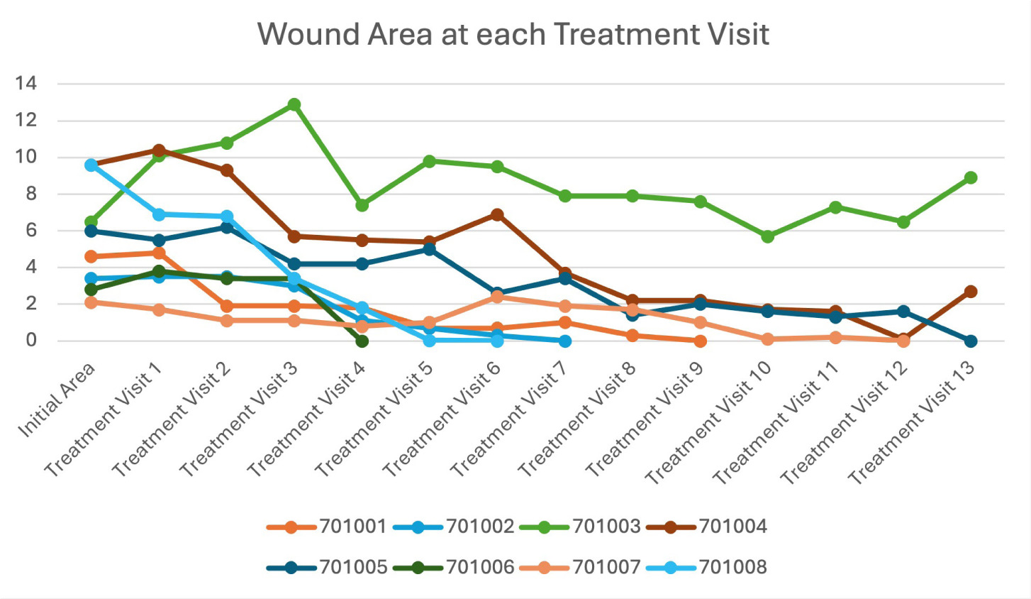
Of the 8 chronic VLU patients in this study population (average initial wound size: 4.96 cm2, SD: 3.29, range: 2.1-9.6 cm2) (Table 2), 6 achieved healing between 3 and 12 applications of NeoThelium FT (75% closure rate) and maintained healed status through the 2-week follow-up period. One patient presented with an initial wound size of 9.60 cm2 (RLE, present for approximately 10 months) and appeared to be on track to heal after achieving a PAR of 98.96% at the 12th treatment visit. However, despite the encouraging progression, the wound grew slightly and ultimately did not reach complete reepithelization with a final PAR of 71.88% after 13 treatment visits and 12 applications. The only patient whose wound failed to improve beyond baseline measurements presented with an initial wound size of 6.50 cm2 that grew to 10.10 cm2 by the first treatment visit—at which point they received the first allograft application. The wound grew to a maximum measurement of 12.9 cm2 by the third treatment visit before displaying a general downward trend in area, reaching a final size of 8.9 cm2. Notably, one of the patients that successfully closed their lesion after 8 applications of NeoThelium FT did so for a VLU on the left lower extremity that had been present for 7 years. This patient had previously failed treatment for multiple advanced interventions, including both CAMPs and a split-thickness skin graft (Figures 1 and 2).
TABLE 2 Average surface area and percent area reduction (PAR) per application
| Total patients | Average area mean ± SD | Median (IQR) | PAR (%) | |
|---|---|---|---|---|
| Initial area | 8 | 4.96 ± 3.29 | 5.30 (3.25–7.28) | N/A |
| Application #1 | 8 | 5.84 ± 3.51 | 5.15 (3.73–7.70) | -17.8 |
| Application #2 | 8 | 4.78 ± 3.72 | 4.85 (3.03–7.43) | 3.59 |
| Application #3 | 8 | 3.96 ± 3.75 | 3.40 (2.73–4.58) | 20.18 |
| Application #4 | 8 | 2.51 ± 2.59 | 1.80 (1.03–4.53) | 49.33 |
| Application #5 | 8 | 2.51 ± 3.55 | 1.00 (0.53–5.10) | 49.26 |
| Application #6 | 8 | 2.80 ± 3.55 | 1.55 (0.23–3.68) | 43.5 |
| Application #7 | 8 | 2.24 ± 2.73 | 1.45 (0.00–3.48) | 54.85 |
| Application #8 | 8 | 1.69 ± 2.66 | 0.85 (0.00–1.83) | 65.95 |
| Application #9 | 8 | 1.60 ± 2.59 | 0.50 (0.00–2.05) | 67.71 |
| Application #10 | 8 | 1.14 ± 1.99 | 0.05 (0.00–1.63) | 77.05 |
| Application #11 | 8 | 1.30 ± 2.51 | 0.10 (0.00–1.38) | 73.77 |
| Application #12 | 8 | 1.03 ± 2.28 | 0.00 (0.00–0.48) | 79.32 |
| Average number of applications received mean ± SD, median | 8.63 ± 3.62, 9.50 | |||
FIGURE 1 Graphical representation of percent area reduction of each patient at each treatment visit
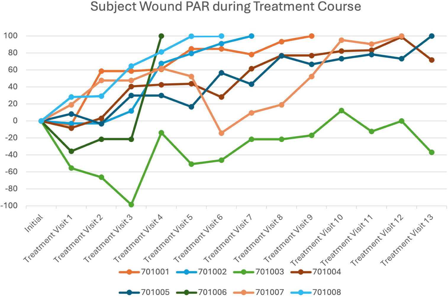
FIGURE 2 Graphical representation of wound area of each patient at each treatment visit
On study completion, after 12 applications the PAR across the study population calculated from aggregate wound area, was 79.32%. The average number of applications required was 8.63 (range 3-12) with a median of 9.5 applications (Table 2) over an average of 9.63 treatment visits. Among the 6 patients that successfully healed, the average number of applications was 7.5 across 8.5 treatment visits. Only two participants displayed an adverse event throughout the course of treatment, neither of which were related to the treated wound or the investigational product, with no serious adverse events being reported. All patients were noted to have tolerated treatment well.
Discussion
In the current healthcare landscape, VLUs constitute an expanding issue, with the dual burden of high prevalence and chronicity. Although helpful, longstanding conventional therapies in VLU treatment fall short in their ability to promote tissue regeneration. The nature of complicated, chronic wounds of venous origin necessitates the incorporation of advanced therapeutic interventions that hold the potential of enhancing wound progression through dermal reconstruction.
The need for novel modalities for VLU treatment—and chronic wounds of other etiological origin—has spurred particular interest in allografts derived from placental tissue. These products have been shown to be composed of various growth factors, structural proteins, chemokines, and cytokines that render them particularly beneficial for chronic wound treatment.17-19 Growth factors native to the tissue, such as fibroblast growth factor, keratinocyte growth factor, vascular endothelial growth factor, and epidermal growth factor, promote the recruitment and proliferation of cells within the wound bed.17,20,32-37 Structural proteins, to include fibronectin, elastin, laminin, and collagens type I-VI, offer a biological scaffolding, enhancing both infiltration and adherence of the recruited and proliferating cells.18 An immunomodulatory effect is exhibited by various cytokines and chemokines in placental tissue, including but not limited to IL-4, IL-6, IL-8, and IL-10, which influence inflammatory responses and contribute to a low risk of graft rejection.17,19,38-41 Thus, placental allografts have exhibited angiogenic/antiangiogenic (tissue dependent), anti-inflammatory, antifibrotic, and antimicrobial effects when applied as a therapeutic intervention for chronic wounds.17,32-37
Particularly high levels of the aforementioned factors native to placental tissue are present in allografts that preserve the amniotic, intermediate/spongy, and chorionic layers in a contiguous state—each layer possesses a unique protein profile in both type and quantity.17-19,42 In other words, products that fit broadly into the category of full-thickness, such as NeoThelium FT, retain the intermediate layer and therefore may preserve more native structure protein content than dual-layer products that separate and recombine layers.27 Interestingly, the contribution of the intermediate/spongy layer, also known as the zona spongiosa, of placental tissue is often absent in commercially available grafts. Amnion-only products by their nature necessitate the removal of both the spongy and chorionic layers during production. Many multi-layered grafts that include the chorion often also lack the zona spongiosa, or include a highly reduced remnant, due to the initial separation and recombination of layers during manufacturing. Noteworthy is the fact that the intermediate layer has been shown to contain over 900 regulatory and signaling components, with an especially high content of hyaluronic acid (HA). This glycosaminoglycan has been associated with analgesic, antifibrotic, and anti-inflammatory effects, which may be of particular interest for VLU treatment.19,42
To fully consider how placental allografts may be a beneficial therapeutic intervention for VLUs, the underlying pathophysiology and pathology of these wounds must be considered. Most venous wounds are a result of chronic vascular insufficiency, a clinically advanced manifestation of chronic venous disease. CVD is a disorder characterized by compromised venous return—the flow of deoxygenated blood back to the heart from the periphery.3,8,10,11 Under normal conditions, this return is driven by musculovenous pumps, also known as peripheral hearts or skeletal muscle pumps. Mechanistically, these pumps function by squeezing local veins when the surrounding muscle contracts. Within these veins are intraluminal, bicuspid valves, which are composed of continuations of the innermost lining of the vein, known as the tunica intima. On muscle contraction, these valves open, allowing prograde flow. When the relevant muscle is in a relaxed state, the valves close, preventing venous reflux. Dysfunction in these pumps from either damage to the intraluminal valves—resulting in reflux—or obstruction from clotting can lead to venous hypertension. The cascade of events initiated by venous hypertension promotes a chronic inflammatory state and edema, ultimately resulting in vein wall and dermal fibrosis, tissue breakdown, and the formation of a painful ulcer.4,5,43 Given the underlying causative factors of these wounds and the associated inflammation, fibrosis, and pain, an intervention shown to have anti-inflammatory, anti-fibrotic, and analgesic effects that can be incorporated into traditional treatment plans may be advantageous.
Irrespective of placental tissue’s promising characteristics for VLU treatment, an insufficient amount of research has been performed regarding the incorporation of placental grafts into treatment protocols. The effectiveness of these products has been well established for DFUs, with one meta-analysis finding that a higher proportion of patients healed at 6 weeks (3.88 times higher) and 12 weeks (2.01 times higher) when treated with a placental graft compared to controls.44 Despite these positive outcomes, inadequate clinical data has been produced to elucidate how effective these products may be for wounds of venous origin.
Outcomes in this small cohort were broadly consistent with those reported for placental allografts in randomized trials, and with the one published full-thickness series.27 The observed 75% closure rate also sits within ranges reported for compression alone in large trials.45,46 However, without complete baseline duration for every ulcer, a formal Morgolis-predicted SOC healing probability could not be calculated for the cohort.47 This study was single-arm and was not powered to detect differences between treatments; the closure rate observed here falls within the range reported for compression therapy alone in large, randomized trials, and the design cannot separate the contribution of the allograft from that of concurrent compression and debridement. These results should therefore be read as clinical experience rather than as evidence of incremental efficacy.
Limitations
Several limitations exist in the presented study that must be considered when interpreting the results. Although appropriate for an investigation of this nature, the most significant limitation was the limited sample size (n=8), which did not allow for advanced statistical analysis and may inhibit reproducibility in future investigations. Additionally, no control group was incorporated, which, although again appropriate for a prospective case series, inhibits the explication of what role the experimental product had in healing compared to SOC. Furthermore, heterogeneity in wound size, severity of pathophysiological mechanisms, past recurrence, and demographics may have exhibited an effect on the results. Despite these limitations, the observations recorded in this case series support further evaluation of NeoThelium FT in controlled studies.
Wound area was estimated by the conventional ruler method (greatest length x perpendicular width). This approach has demonstrated high intra- and intertester reliability and remains widely used in clinical practice and many trials; however, it systemically overestimates true surface area relative to tracing- or image-based planimetry, particularly for irregular wounds.48 Digital planimetry was not used in the present study, so area-change estimates should be interpreted as reliable linear approximations rather than gold-standard planimetric areas. In addition, complete closure was determined by the treating clinician, who was not blinded to treatment allocation. Unblinded clinical assessment of closure introduces a potential risk of detection bias. A subsequent study is necessary to incorporate calibrated digital planimetry for area and independent, blinded adjudication of closure (standardized photographs and predefined epithelialization criteria) to strengthen measurement precision and reduce observer bias.
Conclusion
In this prospective single-arm study, weekly application of a full-thickness placental allograft alongside standard care was well tolerated, with no product-related adverse events and no serious adverse events. Wound progression was observed in 7 of 8 patients, with 6 achieving closure that was maintained through the follow-up period. These observations add to the limited body of clinical data on full-thickness placental allografts in venous leg ulcers and support evaluation of this product in adequately powered controlled studies.