Introduction
Chronic wounds represent a substantial and growing clinical and public health burden. They encompass a heterogeneous group of wounds that fail to progress through an orderly and timely reparative process, including diabetic foot ulcers (DFUs), venous leg ulcers (VLUs), pressure injuries, and other non-healing surgical or traumatic wounds. In the United States, an analysis of Medicare beneficiaries estimated that approximately 8.2 million people had at least one type of wound or wound-related infection, with Medicare expenditure estimates ranging from $28.1 billion to $96.8 billion depending on the wound definition and treatment setting.1 Chronic wounds are associated with prolonged morbidity, recurrent infection, impaired mobility and quality of life, and substantial healthcare utilization.1
DFUs are of particular concern because of their association with infection, recurrence, amputation, and mortality. Approximately 18.6 million people worldwide develop a DFU each year, including an estimated 1.6 million people in the United States.2 Approximately 50–60% of DFUs become infected, and approximately 20% of moderate-to-severe diabetic foot infections result in lower-extremity amputation.2 DFUs precede approximately 80% of lower-extremity amputations among people with diabetes, while recurrence after healing is estimated at 42% at 1 year and 65% at 5 years.2 These outcomes underscore the importance of interventions that address both the underlying etiology of the wound and local factors that impede healing.
Effective wound management requires correction or optimization of the underlying cause wherever possible, together with appropriate local wound care. Contemporary wound-bed preparation involves a systematic approach to optimizing the wound environment through comprehensive assessment, management of the underlying cause, appropriate debridement, control of infection and inflammation, and maintenance of moisture balance.3 Etiology-specific management is also required, including pressure offloading for DFUs and pressure injuries, compression therapy for appropriate VLUs, optimization of tissue perfusion, infection management, and management of relevant systemic factors.2,3
Debridement is an important component of wound-bed preparation because devitalized tissue, slough, debris, and microbial biofilm can impede wound assessment and create barriers to repair.3 Biofilm is particularly relevant in chronic wounds because microorganisms embedded within a biofilm phenotype demonstrate increased tolerance to host defenses and antimicrobial interventions and may contribute to persistent inflammation and delayed healing. A systematic review and meta-analysis estimated that biofilm was present in approximately 78% of chronic wounds, although substantial methodological heterogeneity in biofilm detection was noted.4 Removal or disruption of these barriers is therefore an important consideration in wound-bed management.3,4
Sharp and surgical debridement permit rapid removal of devitalized tissue, but no single debridement modality is appropriate for every wound or patient. Selection depends on wound characteristics, treatment objectives, perfusion, pain and tolerance, bleeding risk, clinician expertise, and the clinical setting.3 Evidence comparing debridement approaches is also limited; for example, a Cochrane review of debridement for VLUs identified considerable heterogeneity and insufficient evidence to establish the superiority of a particular approach.5 These considerations have supported continued investigation of adjunctive technologies capable of facilitating selective removal of nonviable tissue and wound-bed preparation.
Low-frequency ultrasound (LFU) has emerged as one such modality. LFU systems used in wound care typically operate at frequencies substantially below those used for diagnostic ultrasound and may employ either contact or noncontact delivery.6 Acoustic mechanisms associated with LFU include cavitation and acoustic microstreaming, which can produce mechanical effects within the wound environment and facilitate disruption and removal of devitalized tissue and biofilm.6 The clinical literature has evaluated LFU across multiple chronic wound etiologies using different devices, frequencies, intensities, and treatment protocols. A systematic review of LFU debridement identified potential benefits including reductions in slough and exudate, biofilm disruption, pain reduction, and improved wound healing, but emphasized that much of the available evidence was derived from relatively small and methodologically heterogeneous studies.6 A systematic review and meta-analysis of eight randomized controlled trials similarly found evidence supporting LFU as an adjunct to standard wound management, while highlighting heterogeneity across treatment modalities and study designs.7
The Qoustic Wound Therapy System™ (QWTS; Arobella Medical, LLC, Minnetonka, MN, USA) is a low-frequency ultrasound system designed for contact and noncontact wound treatment. The system operates at a nominal frequency of 35 kHz (±3 kHz) and uses a proprietary dome-shaped Qoustic Qurette™ applicator with an irrigation medium. Its cleared indications include selective and nonselective dissection and fragmentation of soft or hard tissue; surgical, excisional, or sharp-edge debridement of acute and chronic wounds and burns; cleansing irrigation and lavage; contact or noncontact maintenance debridement; and wound-bed preparation for grafting or other subsequent procedures. The system therefore combines low-frequency ultrasonic energy and irrigation with treatment modes that permit both contact debridement and noncontact application.8,9
QWTS-specific preclinical studies provide mechanistic evidence relevant to wound management. Karau et al. evaluated the system against planktonic and biofilm forms of Pseudomonas aeruginosa, Staphylococcus epidermidis, and Staphylococcus aureus in vitro.10 Four minutes of treatment reduced planktonic bacterial concentrations by mean values of 5.10, 4.99, and 5.22 log10 colony-forming units/mL, respectively, while 10 minutes of treatment reduced established biofilms by 1.34, 1.46, and 1.02 log10 colony-forming units/cm2, respectively.10 These findings demonstrated antibacterial and biofilm-reducing effects under controlled in vitro conditions but do not, in isolation, establish clinical effectiveness. Additional in vitro work examining 35-kHz LFU exposure in human fibroblasts identified changes in fibroblast morphology and migration patterns compared with untreated controls, providing further evidence that LFU may influence cellular processes relevant to tissue repair.11
Despite these mechanistic findings and the broader clinical literature examining LFU in wound management, evidence specific to QWTS in routine clinical practice remains limited. Importantly, findings obtained with other LFU systems cannot necessarily be extrapolated directly to QWTS because ultrasound devices differ in frequency, intensity, delivery method, applicator design, contact with the wound, and treatment protocol.6 Evidence evaluating QWTS across heterogeneous wound etiologies in multicenter outpatient clinical practice is also limited.
The present retrospective multicenter study therefore evaluated wound-healing outcomes among patients treated with QWTS in conjunction with standard of care (SOC) in private outpatient wound medicine practices. The primary objective was to characterize cumulative complete wound closure over time across DFUs, VLUs, pressure injuries, and open wounds. The secondary objective was to evaluate percent area reduction (PAR) among treated wounds. By examining routinely collected longitudinal clinical data, the study sought to characterize real-world wound-healing trajectories during QWTS treatment across heterogeneous wound etiologies.
Methods
Study design and setting
This retrospective, multicenter study evaluated real-world clinical outcomes among patients with full-thickness wounds treated with the Qoustic Wound Therapy System as an adjunct to SOC. The study was conducted across private outpatient wound medicine practices in Louisiana, Mississippi, Arkansas, Tennessee, and Texas. All treatments and clinical assessments were performed as part of routine outpatient wound care, and the analysis was based on retrospectively collected billing and electronic health record (EHR) data.
Study population and case identification
The study population comprised patients with full-thickness wounds who received QWTS treatment at participating outpatient wound medicine practices during the study period. Patients were initially identified from accounts receivable reports using Current Procedural Terminology (CPT) code 97610, which denotes low-frequency, noncontact, nonthermal ultrasound treatment, including topical application(s), wound assessment, and instructions for ongoing care, per day.
Following identification of QWTS-treated patients, wound-level clinical information was obtained from the EHR. Wound etiology was determined using International Classification of Diseases, Tenth Revision (ICD-10) diagnosis codes documented in the accounts receivable reports and EHR and subsequently classified as DFU, VLU, pressure injury, or open wound according to prespecified clinical definitions. Because the open-wound category encompassed several clinically distinct wound types, its constituent etiologies were additionally characterized descriptively as part of a post hoc analysis. Patient demographic and wound characteristics were abstracted manually from the EHR by a single investigator.
For descriptive characterization of the heterogeneous open-wound cohort, the constituent wound types were additionally grouped post hoc according to the pathophysiologic categories: microvascular/ischemic wounds (arterial ulcers and failed surgical grafts), acute mechanical/thermal wounds (trauma, open surgical wounds, and burns), and miscellaneous/unclassified wounds (Other). These groupings were used for descriptive purposes only and were not incorporated as separate categories in the primary Bayesian model.
Because CPT 97610 is reported once per treatment encounter rather than separately for each wound, billing data could not independently identify every wound receiving QWTS when multiple wounds were treated during the same encounter. For patients with multiple wounds, the presence of CPT 97610 therefore established that QWTS treatment occurred during the encounter but did not permit definitive attribution of treatment to each individual wound in the absence of corresponding wound-level documentation.
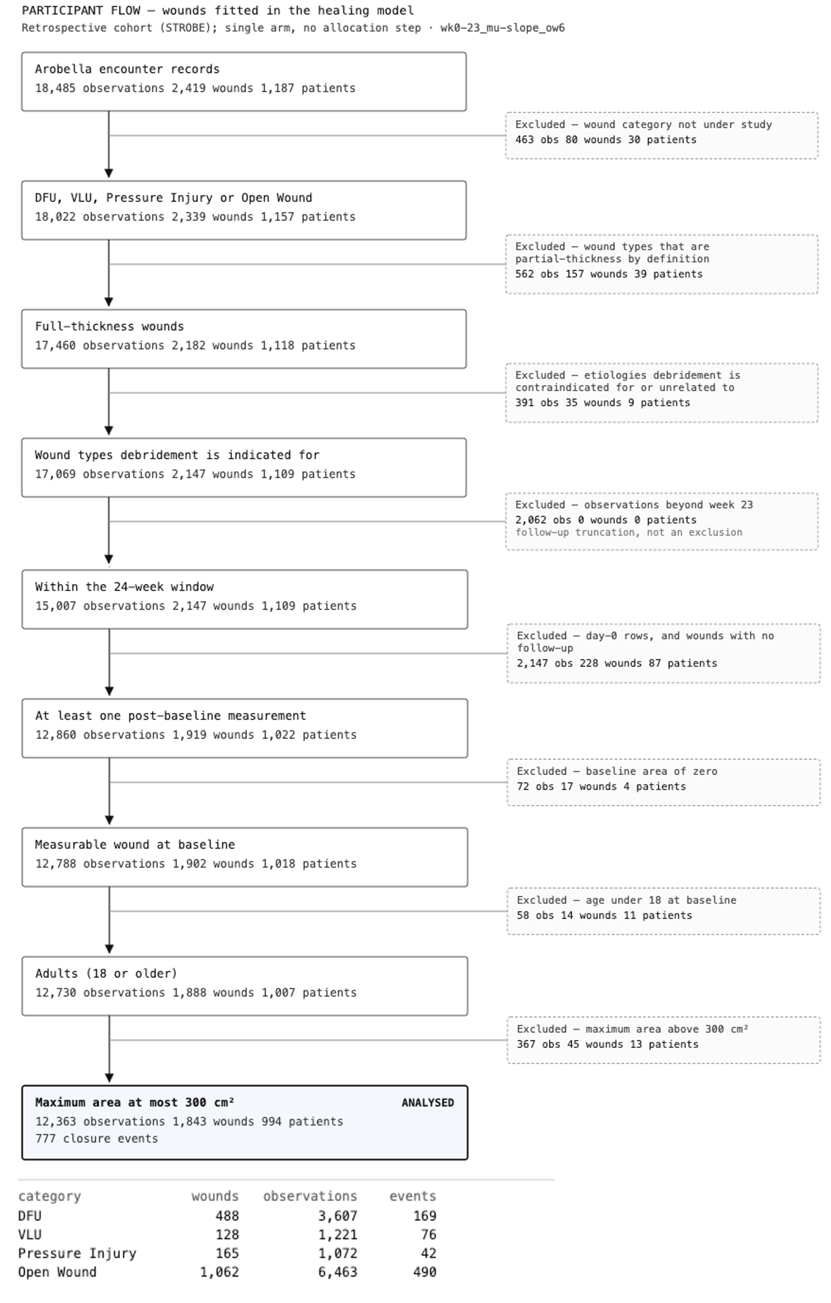
Eligible wounds were required to be full-thickness wounds within the prespecified wound categories, with a measurable baseline wound area and at least one post-baseline measurement. Patients were required to be aged ≥18 years. Wounds with a maximum observed area >300 cm2 were excluded, and longitudinal observations beyond week 23 were excluded from the analytic dataset. Partial-thickness wound types and wound etiologies for which ultrasonic debridement was considered contraindicated or unrelated to the underlying wound mechanism were excluded. The derivation of the analytic cohort is shown in Figure 1.
FIGURE 1 Derivation of the analytic cohort for the wound-healing model..
Wound classification
Wounds were classified as DFUs, VLUs, pressure injuries, or open wounds. DFUs were defined as full-thickness wounds located on the ankle or foot in patients with a confirmed diagnosis of type 1 or type 2 diabetes mellitus. VLUs were defined as full-thickness wounds of the lower extremity in patients with venous insufficiency confirmed by duplex ultrasound demonstrating venous reflux greater than 0.5 seconds. Pressure injuries were defined as full-thickness wounds occurring over a bony prominence or attributable to pressure from a medical device, irrespective of anatomical location. Open wounds comprised full-thickness wounds that did not meet the criteria for classification as a DFU, VLU, or pressure injury and for which ultrasonic debridement was considered clinically applicable. This category included wounds classified as Other, Trauma, Surgical Wound–Open, Arterial Ulcer, Burn, and Failed Surgical Graft. Skin tears, abrasions, and ruptured blisters were excluded as partial-thickness wounds. Autoimmune wounds, pyoderma, radiation injuries, and carcinoma-associated wounds were excluded because ultrasonic debridement was considered contraindicated or not relevant to the underlying wound mechanism.
Qoustic Wound Therapy System and treatment
QWTS is a low-frequency ultrasound system operating at a nominal frequency of 35 kHz (±3 kHz) and uses a Qoustic Qurette applicator with an irrigation medium. The system permits both contact and noncontact treatment and can be used for wound debridement, cleansing and irrigation, maintenance debridement, and wound-bed preparation for subsequent procedures.
For the present study, QWTS was used to provide low-frequency, noncontact, nonthermal ultrasound treatment. Treating clinicians followed institutional protocols and used the system in accordance with the manufacturer’s Instructions for Use and applicable Local Coverage Determination guidance. Treatment was delivered with continuous irrigation, as required for operation of the system. Each treatment used a Qoustic Qurette Disposable Tubing Set, a proprietary system accessory that connects the irrigation-fluid reservoir to the Qoustic Qurette applicator and provides fluid-flow control during treatment. The tubing set is designated for single-patient, single-treatment use and was disposed of after treatment in accordance with the manufacturer’s Instructions for Use. Permitted irrigation media include sterile 0.9% saline, sterile de-ionized water, Vashe™ Wound Cleanser, or other solutions approved for wound therapy or debridement.
During noncontact treatment, the Qoustic Qurette may be positioned with light guidance contact or held approximately 1–2 cm above the wound surface, with the applicator moved continuously over the treatment area. Recommended treatment duration is based on treatment area, ranging from 1 minute for wounds measuring <40 cm2 to 5 minutes for wounds measuring 160–200 cm2.
Patients were scheduled for weekly visits, during which QWTS treatment was performed as clinically indicated. A treatment series was defined as consecutive QWTS treatments with no more than 14 days between successive treatments. Treatment continued until completion of the treatment series or achievement of complete wound closure.
Standard of care
QWTS was administered in conjunction with SOC. Concurrent SOC included wound debridement, confirmation of adequate perfusion, management of infection and necrotic tissue, and appropriate offloading or compression therapy according to wound etiology and clinical indication. Nutritional status was assessed, with counseling or intervention provided when indicated. Smoking-cessation counseling was offered where appropriate. Patients with poorly controlled diabetes received targeted education, medication adjustment, and referral to primary care or endocrinology as needed. Dressings were selected at the treating clinician’s discretion according to wound characteristics, including the need to manage exudate and minimize bioburden accumulation.
Wound assessment and measurement
Wound surface area was measured in square centimeters (cm2) using a standardized ruler-based length × width method at all participating sites. Baseline wound area was defined as the pre-debridement measurement obtained at the first QWTS treatment. Thereafter, wound measurements obtained during longitudinal follow-up were incorporated into the analysis, allowing wound status and time-varying covariates to be updated across longitudinal observations.
Complete wound closure was defined as a 99.99–100% reduction in wound area from baseline. This threshold was applied because clinicians occasionally documented a residual wound area of 0.01 cm2 before subsequent follow-up and discharge, at which point wound surface area was documented as 0.0 cm2.
Data variables
Patients could contribute more than one wound to the analysis. No minimum baseline wound-area threshold was applied to the primary analytic population. To maintain de-identification in accordance with the Health Insurance Portability and Accountability Act (HIPAA) Safe Harbor provisions, age was capped at 90 years. Sex and ethnicity were reported as recorded administratively in the source data. Race was not captured in the source extract and was therefore unavailable for analysis. Diabetes status reflected the presence of a diabetes diagnosis code in the available records; absence of such a code was not interpreted as confirmation that diabetes was absent.
Outcomes
The primary outcome was cumulative complete wound closure over the observation period following treatment with QWTS in conjunction with SOC. Model-standardized cumulative closure probabilities were estimated at weeks 4, 8, 12, 16, and 20. The secondary outcome was percent area reduction (PAR) from baseline, with week 12 prespecified as the primary comparison time point.
Statistical analysis
Analyses were conducted within a Bayesian longitudinal framework using a hurdle-gamma model for repeated wound-area measurements. The model comprised two components: a hurdle component modeling the probability that wound area remained greater than zero (i.e., the wound had not achieved complete closure) using a logit link, and a gamma component modeling mean wound area among observations with positive wound area using a log link. A common gamma shape parameter was estimated across observations. Because the hurdle component modeled the probability of remaining open, positive coefficients in this component corresponded to a lower probability of closure. The four prespecified wound categories were retained in the primary model. The post hoc open-wound subgroups were not modeled separately because the microvascular/ischemic subgroup contained only 9 closure events among 46 wounds, providing insufficient information for reliable estimation of a category-specific model term.
Both model components included category-specific intercepts and week slopes and adjusted for log-transformed baseline wound area centered on the mean of the analytic population. Patient-level random intercepts were included in both components, as were wound-level random intercepts, with a wound-level random week slope included in the positive-area component. Random effects were specified using a non-centered parameterization; wound-level offsets were constrained to sum to zero within wound category and patient-level offsets to sum to zero across patients. The model was fitted to 12,363 longitudinal observations from 1,843 wounds among 994 patients, including 777 closure events.
Weakly informative priors were specified for all model parameters. For the hurdle component, category-specific intercepts were assigned Normal(2.9, 1.0) priors, week effects Normal(0, 0.3) priors, and the coefficient for log baseline wound area a Normal(0, 0.5) prior. Patient- and wound-level standard deviations were assigned HalfNormal(0.75) priors. For the positive wound-area component, category-specific intercepts were assigned Normal(0.65, 0.75) priors, week effects Normal(0, 0.15) priors, and the coefficient for log baseline wound area a Normal(0.5, 0.4) prior. Patient- and wound-level intercept standard deviations were assigned HalfNormal(0.5) priors, and the wound-level week-slope standard deviation was assigned a HalfNormal(0.2) prior. The gamma shape parameter was assigned a Gamma(4.0, 1.0) prior.
Posterior distributions were estimated using Markov chain Monte Carlo sampling with the No-U-Turn Sampler (NUTS). Four chains were run, each with 1,000 tuning iterations followed by 4,000 posterior draws, yielding 16,000 post-tuning draws. The target acceptance probability was 0.90. Convergence and sampling adequacy were assessed using R-hat statistics, divergent transitions, and bulk and tail effective sample sizes. Prespecified criteria were R-hat ≤1.01, no divergent transitions, and effective sample sizes ≥400; all criteria were met. Posterior uncertainty was summarized using 95% equal-tailed credible intervals.
Model-standardized cumulative closure probabilities were estimated using g-computation over the empirical covariate distribution. Each wound contributed its observed covariates and random effects, and predicted probabilities were averaged across wounds within each posterior draw. Standardized cumulative closure trajectories were estimated at weeks 4, 8, 12, 16, and 20 assuming weekly observations. The model did not include a treatment dose or treatment-frequency term; consequently, the standardized observation schedule determined the modeled timing of wound assessment and should not be interpreted as a counterfactual intervention assigning weekly QWTS treatment.
Percent area reduction (PAR) was estimated from the fitted Bayesian hurdle-gamma model as the reduction in total expected wound area at each evaluated week relative to total baseline wound area. Estimates were standardized over the analysis population, with each wound evaluated at its own baseline area and estimated patient- and wound-level effects. Wounds classified as completely closed contributed an area of zero. PAR estimates were summarized as posterior means with 95% equal-tailed credible intervals. Week 12 was prespecified as the primary comparison time point for PAR.
No missing values were imputed. Structural null values arising from the data structure were distinguished from genuine missing observations. Baseline covariates included in the fitted model were complete within the analytic population; therefore, no complete-case restriction or covariate imputation was required for model estimation. Comorbidity indicators represented whether a condition had ever been coded in the available records; an unflagged condition was not interpreted as confirmed absence of that condition.
Analyses were performed in Python version 3.14.6 using PyMC version 6.2.0, with NUTS implemented through nutpie version 0.16.11. ArviZ version 1.2.0 was used for posterior diagnostics.
Ethical considerations
The study protocol, Wound Care Outcomes via EHR for Retrospective Research, was reviewed by Sterling IRB (IRB ID 17333-AFerguson) under an exempt review. A waiver of authorization for the retrospective use of protected health information was approved on July 30, 2026. The waiver was granted on the basis that the use or disclosure of protected health information involved no more than minimal risk to individuals, the research could not practicably be conducted without the waiver and access to the relevant protected health information, and appropriate safeguards were in place to protect privacy and confidentiality. The study was conducted in accordance with the ethical standards of the responsible institutional review board and with the principles of the Declaration of Helsinki.
Results
Study population and baseline characteristics
The derivation of the analytic cohort is shown in Figure 1. The final analytic population comprised 994 patients contributing 1,843 wounds and 12,363 longitudinal wound observations. The cohort included 488 DFUs, 128 VLUs, 165 pressure injuries, and 1,062 open wounds.
Detailed baseline characteristics were available for the DFU, VLU, and pressure injury cohorts, comprising 457 patients and 781 wounds. Of these patients, 172 (37.6%) contributed more than one wound. Median age at baseline was 65 years (IQR, 54–75; range, 24–90), 318 (69.6%) were male and 139 (30.4%) were female, and a diabetes diagnosis was recorded for 349 (76.4%).
Of the 781 wounds, 488 (62.5%) were DFUs, 128 (16.4%) were VLUs, and 165 (21.1%) were pressure injuries. Median baseline wound area was 1.7 cm2 (IQR, 0.4–7.9; range, 0.01–283.5) overall. Baseline wound area varied by wound etiology, with median areas of 1.2 cm2 (IQR, 0.3–5.2) for DFUs, 3.3 cm2 (IQR, 1.0–14.6) for VLUs, and 2.3 cm2 (IQR, 0.6–11.6) for pressure injuries. Overall, 312 wounds (39.9%) had a baseline area <1.00 cm2. Wounds contributed a median of 4 longitudinal observations (IQR, 2–11; range, 1–45), with a median last observed week of 4 (IQR, 2–10; range, 0–23). Baseline patient and wound characteristics are summarized in Table 1.
TABLE 1 Baseline patient and wound characteristics of the DFU, VLU, and pressure injury cohorts
| Characteristic | DFU | VLU | Pressure injury | Overall |
|---|---|---|---|---|
| Patients (n=457) | ||||
| Age at baseline, years, | 65.0 | |||
| Median [IQR] | [54.0–75.0] | |||
| (Range) | (24.0–90.0) | |||
| At age cap of 90, n (%) | — | — | — | 12 (2.6) |
| Female, n (%) | — | — | — | 139 (30.4) |
| Male, n (%) | — | — | — | 318 (69.6) |
| Not Hispanic or Latino, n (%) | — | — | — | 345 (75.5) |
| Declined to specify ethnicity, n (%) | — | — | — | 100 (21.9) |
| Ethnicity not recorded, n (%) | — | — | — | 6 (1.3) |
| Hispanic or Latino, n (%) | — | — | — | 4 (0.9) |
| Refused to report ethnicity, n (%) | — | — | — | 2 (0.4) |
| Diabetes diagnosis coded, n (%) | — | — | — | 349 (76.4) |
| Contributing >1 wound, n (%) | — | — | — | 172 (37.6) |
| Wounds (n=781) | ||||
| Wounds, n (%) | 488 (62.5) | 128 (16.4) | 165 (21.1) | 781 (100.0) |
| Baseline area, cm2, median | 1.2 | 3.3 | 2.3 | 1.7 |
| [IQR] | [0.3–5.2] | [1.0–14.6] | [0.6–11.6] | [0.4–7.9] |
| (range) | (0.01–229.5) | (0.06–283.5) | (0.01–174.2) | (0.01–283.5) |
| Maximum observed wound area during follow-up, | 2.2 | 8.3 | 4.0 | 2.9 |
| cm2, median [IQR] | [0.5–7.5] | [2.3–27.6] | [0.9–15.4] | [0.7–11.5] |
| (range) | (0.01–229.5) | (0.06–283.5) | (0.01–224.0) | (0.01–283.5) |
| Observations per wound, median | 4 | 6 | 4 | 4 |
| [IQR] | [2–10] | [2–15] | [2–8] | [2–11] |
| (range) | (1–44) | (1–42) | (1–45) | (1–45) |
| Last observed follow-up week, median | 3 | 5 | 3 | 4 |
| [IQR] | [2–9] | [2–13] | [2–10] | [2–10] |
| (range) | (0–23) | (0–23) | (0–23) | (0–23) |
| Baseline area <1.00 cm2, n (%) | 223 (45.7) | 32 (25.0) | 57 (34.5) | 312 (39.9) |
Abbreviations: DFU, diabetic foot ulcer; IQR, interquartile range; VLU, venous leg ulcer. Notes: Age was capped at 90 years in accordance with HIPAA Safe Harbor requirements. Sex and ethnicity reflect administrative recording in the source data. Race was not captured and is therefore not reported. “Diabetes diagnosis coded” indicates that a diabetes diagnosis was identified in the available records; absence of a diabetes code does not establish absence of diabetes. Patients could contribute more than one wound; consequently, patient- and wound-level denominators differ. No minimum baseline wound-area threshold was applied to this analytic population. Detailed baseline characteristics for the open-wound cohort were not included in this descriptive baseline dataset.
Open-wound cohort
The open-wound category represented a heterogeneous group of full-thickness wounds retained on clinical grounds. It comprised Other (n=460), Trauma (n=355), Surgical Wound–Open (n=160), Arterial Ulcer (n=43), Burn (n=41), and Failed Surgical Graft (n=3). Autoimmune wounds, pyoderma, radiation injuries, and carcinoma-associated wounds were excluded from this analytic category. In the post hoc descriptive grouping, 556 wounds (52.4% of the open-wound cohort) were classified as acute mechanical/thermal wounds, comprising trauma, open surgical wounds, and burns; 46 (4.3%) were classified as microvascular/ischemic wounds, comprising arterial ulcers and failed surgical grafts; and 460 (43.3%) were classified as miscellaneous/unclassified wounds. Baseline wound area and crude observed closure characteristics for these subgroups are presented in Table 2. These descriptive closure rates are unadjusted for follow-up duration and should not be interpreted as equivalent to the model-standardized closure probabilities reported in the primary analysis.
TABLE 2 Descriptive characteristics of the open-wound cohort and post hoc clinical subgroups
| Open-wound group | Wounds, n (%) | Patients, n | Observations, n | Baseline area, cm2, median [IQR] | Wounds closing at least once, n (%) |
|---|---|---|---|---|---|
| Acute mechanical/thermal | 556 (52.4) | 369 | 3,211 | 2.5 [0.8–8.8] | 242 (43.5) |
| Microvascular/ischemic | 46 (4.3) | 29 | 445 | 3.4 [1.2–13.8] | 9 (19.6) |
| Miscellaneous/unclassified | 460 (43.3) | 250 | 2,807 | 2.9 [0.5–11.1] | 192 (41.7) |
Acute mechanical/thermal wounds comprised trauma, open surgical wounds, and burns; microvascular/ischemic wounds comprised arterial ulcers and failed surgical grafts; miscellaneous/unclassified wounds comprised wounds recorded as Other. Values are descriptive observed data and are not model-standardized estimates.
Model-standardized wound closure
Model-standardized cumulative closure probabilities increased progressively across the 20-week standardized observation period. Across all 1,843 wounds included in the four-category analysis, the estimated cumulative probability of complete wound closure was 29.3% (95% CrI, 27.9%–30.7%) by week 4, 41.5% (95% CrI, 39.7%– 43.3%) by week 8, 49.4% (95% CrI, 47.3%–51.5%) by week 12, 55.2% (95% CrI, 52.7%–57.7%) by week 16, and 59.8% (95% CrI, 56.8%–62.8%) by week 20.
Model-standardized closure trajectories showed variation across wound etiologies. At week 12, the estimated cumulative probability of complete closure was 41.8% (95% CrI, 38.0%–45.7%) for DFUs, 50.5% (95% CrI, 45.0%– 56.2%) for VLUs, 35.8% (95% CrI, 29.4%–42.3%) for pressure injuries, and 54.8% (95% CrI, 52.1%–57.7%) for open wounds. By week 20, the corresponding probabilities were 53.7% (95% CrI, 48.0%–59.5%), 63.6% (95% CrI, 55.9%– 71.0%), 42.8% (95% CrI, 33.9%–52.5%), and 64.8% (95% CrI, 60.9%–68.8%), respectively. Model-standardized cumulative closure probabilities across all evaluated time points are presented in Table 3.
TABLE 3 Model-standardized cumulative probability of complete wound closure by wound etiology and follow-up week, % (95% CrI)
| Wound group | Wounds, n | Week 4 | Week 8 | Week 12 | Week 16 | Week 20 |
|---|---|---|---|---|---|---|
| All wounds | 1,843 | 29.3 (27.9–30.7) |
41.5 (39.7–43.3) |
49.4 (47.3–51.5) |
55.2 (52.7–57.7) |
59.8 (56.8–62.8) |
| DFU | 488 | 22.3 (19.7–24.9) |
33.6 (30.4–37.0) |
41.8 (38.0–45.7) |
48.3 (43.6–53.0) |
53.7 (48.0–59.5) |
| VLU | 128 | 28.5 (24.1–33.1) |
41.5 (36.5–46.7) |
50.5 (45.0–56.2) |
57.6 (51.3–64.0) |
63.6 (55.9–71.0) |
| Pressure injury | 165 | 21.4 (17.1–26.0) |
30.2 (24.9–35.8) |
35.8 (29.4–42.3) |
39.8 (32.2–47.7) |
42.8 (33.9–52.5) |
| Open wound | 1,062 | 33.9 (32.0–35.9) |
46.9 (44.6–49.2) |
54.8 (52.1–57.7) |
60.5 (57.2–63.9) |
64.8 (60.9–68.8) |
Abbreviations: CrI, credible interval; DFU, diabetic foot ulcer; VLU, venous leg ulcer. Note: Values are model-standardized cumulative closure probabilities with 95% equal-tailed credible intervals. Complete wound closure was defined as a 99.99%–100% reduction in wound area from baseline. Estimates were standardized over the empirical covariate distribution using g-computation, with wounds contributing their observed covariates and random effects. Standardized trajectories assume weekly observations through week 20. The model does not include a treatment dose or treatment-frequency term; consequently, these estimates describe model-standardized closure trajectories under the specified observation schedule and should not be interpreted as counterfactual estimates of the effect of weekly QWTS treatment. Credible intervals represent posterior uncertainty in the population-level mean estimate rather than variation among individual wounds.
Percent area reduction
At the prespecified week-12 comparison, total expected wound area was reduced by an estimated 50.4% (95% CrI, 47.1%–53.3%) from baseline across the overall analysis population. Model-estimated PAR at week 12 was 47.5% (95% CrI, 38.9%–53.4%) for DFUs, 34.6% (95% CrI, 23.0%–42.9%) for VLUs, 33.3% (95% CrI, 20.8%–42.9%) for pressure injuries, and 56.2% (95% CrI, 52.1%–59.7%) for open wounds.
PAR increased progressively across the evaluated time points, from 25.4% (95% CrI, 20.7%–29.3%) at week 4 to 66.8% (95% CrI, 63.9%–69.4%) at week 20 for the overall population. By week 20, estimated PAR was 63.6% (95% CrI, 56.7%–68.8%) for DFUs, 47.2% (95% CrI, 34.5%–56.9%) for VLUs, 53.2% (95% CrI, 42.1%–62.0%) for pressure injuries, and 72.7% (95% CrI, 69.4%–75.6%) for open wounds. Model-estimated PAR across all evaluated time points is presented in Table 4.
TABLE 4 Model-estimated percent area reduction by wound etiology and follow-up week, % (95% CrI)
| Wound group | Wounds, n | Week 4 | Week 8 | Week 12 | Week 16 | Week 20 |
|---|---|---|---|---|---|---|
| All wounds | 1,843 |
25.4 (20.7–29.3) |
39.3 (35.4–42.4) |
50.4 (47.1–53.3) |
59.5 (56.4–62.2) |
66.8 (63.9–69.4) |
| DFU | 488 | 24.3 (12.6–31.8) |
36.9 (27.1–43.5) |
47.5 (38.9–53.4) |
56.2 (48.7–61.7) |
63.6 (56.7–68.8) |
| VLU | 128 | 19.6 (5.9–28.9) |
27.4 (15.3–35.7) |
34.6 (23.0–42.9) |
41.2 (29.1–50.1) |
47.2 (34.5–56.9) |
| Pressure injury | 165 | 4.8 (−11.6–17.6) |
20.3 (6.4–31.0) |
33.3 (20.8–42.9) |
44.1 (32.5–53.3) |
53.2 (42.1–62.0) |
| Open wound | 1,062 | 29.8 (23.9–34.5) |
44.6 (39.8–48.4) |
56.2 (52.1–59.7) |
65.5 (61.8–68.6) |
72.7 (69.4–75.6) |
Abbreviations: CrI, credible interval; DFU, diabetic foot ulcer; PAR, percent area reduction; VLU, venous leg ulcer.
Note: Values are posterior mean percent area reductions with 95% equal-tailed credible intervals. PAR was estimated from the fitted Bayesian hurdle-gamma model as the reduction in total expected wound area relative to total baseline wound area, standardized over the analysis population at each wound’s baseline area and estimated patient- and wound-level effects. Completely closed wounds contributed an area of zero. Week 12 was the prespecified primary comparison time point.
Discussion
In this retrospective multicenter study of 994 patients contributing 1,843 wounds, model-standardized cumulative closure probabilities increased progressively across the 20-week observation period. The estimated cumulative probability of complete closure for the overall cohort was 29.3% by week 4, 49.4% by week 12, and 59.8% by week 20. At the prespecified week-12 comparison, estimated PAR was 50.4% overall, with estimates of 47.5% for DFUs, 34.6% for VLUs, 33.3% for pressure injuries, and 56.2% for open wounds. Together, the closure and PAR analyses characterize both achievement of complete closure and changes in wound area among the treated population.
These findings provide device-specific real-world evidence describing longitudinal wound healing across several clinically distinct wound categories among patients receiving QWTS in conjunction with SOC. They should not, however, be interpreted as estimates of the independent treatment effect of QWTS. The study lacked a concurrent comparator, and the model-standardized analyses did not explicitly model QWTS dose or treatment frequency.
Accordingly, the estimates characterize wound-healing trajectories in the treated population under the specified standardization procedures rather than the causal effect of QWTS.
The present findings are directionally consistent with a body of clinical literature suggesting that low-frequency ultrasound may support healing when used as an adjunct to conventional wound care, but that literature is heterogeneous and does not permit straightforward numerical comparison with the present estimates. The systematic review by Voigt et al. identified eight randomized trials of LFU in DFUs and venous ulcers and reported evidence of improved early healing with both contact and noncontact approaches, while also emphasizing substantial risk of bias, particularly in studies of low-intensity noncontact ultrasound.7 This heterogeneity remains important because ultrasound systems differ in frequency, intensity, delivery technique, treatment schedule, and clinical indication.
For DFUs, some randomized studies of noncontact LFU have reported favorable healing outcomes. In the multicenter, randomized, double-blind, sham-controlled study by Ennis et al, appropriately treated DFUs receiving noncontact kilohertz ultrasound plus SOC had a 12-week healing rate of 40.7%, compared with 14.3% in the sham-treated efficacy group.12 A later randomized, double-blind sham-controlled study by Rastogi et al also found more rapid early wound contraction with airborne LFU, although the difference in overall percentage area reduction at 4 weeks was not statistically significant.13 Yao et al subsequently explored treatment dose and biological response in nonhealing DFUs and demonstrated that treatment frequency may itself influence wound response, reinforcing the importance of exposure intensity when interpreting ultrasound studies.14
The present DFU findings provide complementary information on both wound-area reduction and complete closure. Estimated PAR was 47.5% at the prespecified week-12 comparison and increased to 63.6% by week 20, while the model-standardized cumulative probability of complete closure was 53.7% by week 20. These findings are broadly consistent with prior evidence documenting wound improvement during LFU-assisted care, including studies evaluating percentage area reduction.13 Direct numerical comparison is nevertheless inappropriate because previous studies evaluated different ultrasound devices, treatment protocols, populations, and outcome definitions, whereas the present estimates derive from longitudinal observational data and do not estimate an incremental QWTS effect.
PAR complements complete closure by capturing wound-area change among wounds that had not necessarily achieved closure. PAR increased over time across each wound category, although the magnitude and uncertainty varied by etiology. As with the closure findings, these estimates do not isolate the contribution of QWTS from SOC or other aspects of clinical management.
Evidence for LFU in VLUs is mixed. Randomized studies of noncontact LFU have reported improvements in wound-area reduction relative to SOC in some settings. Olyaie et al randomized 90 patients to SOC, high-frequency ultrasound plus SOC, or noncontact LFU plus SOC and reported greater wound-area reduction with ultrasound-treated groups.15 White et al, however, found that adding noncontact LFU three times weekly to UK SOC did not produce unequivocal superiority across all healing outcomes in a single-center assessor-blinded randomized trial, underscoring the variability of results across settings and treatment protocols.16 Other work using direct-contact LFU debridement has shown faster VLU healing than sharp debridement, but this involved a substantially different 22.5-kHz high-intensity contact system and therefore cannot be considered directly comparable to QWTS.17
Against this background, the present VLU findings provide complementary information on both wound-area reduction and complete closure during QWTS-assisted care. Estimated PAR was 34.6% at the prespecified week-12 comparison and 47.2% by week 20, while the model-standardized cumulative probability of complete closure was 63.6% by week 20. Differences in study design, ultrasound technology, treatment protocols, and outcome definitions preclude direct numerical comparison with previous trials. The present findings should therefore be interpreted descriptively and cannot establish superiority over compression-based SOC or determine the incremental contribution of QWTS.
The pressure-injury literature is less consistent. A multicenter randomized placebo-controlled trial by ter Riet et al found 12-week closure in 40% of ultrasound-treated pressure ulcers compared with 44% of sham-treated ulcers, with essentially no difference in the estimated closure rate between groups.18 More recent retrospective work has explored noncontact LFU specifically in deep-tissue pressure injuries, but the evidence remains observational and comparatively limited.19 The lower week-20 standardized closure probability observed for pressure injuries in the present cohort (42.8%) may reflect differences in underlying pathophysiology, patient characteristics, wound severity, or supportive care; however, these explanations were not evaluated in the present analysis.
Evidence relating to the heterogeneous open-wound category is more difficult to contextualize because there is no directly comparable composite population in the existing LFU literature. Previous studies have evaluated noncontact LFU in heterogeneous chronic lower-extremity wounds and ischemic foot and leg ulcers, with some reporting improved wound-healing outcomes when LFU was incorporated into care.20,21 However, differences in wound classification, ultrasound technology, treatment protocols, comparators, and outcome definitions limit comparison with the present cohort. The 64.8% week-20 estimate observed for open wounds in the present analysis should therefore not be interpreted as a common expected closure probability across its constituent traumatic, surgical, arterial, burn, failed-graft, and other wound subtypes. Post hoc descriptive stratification further demonstrated the composition of this cohort, with acute mechanical/thermal wounds accounting for 52.4%, miscellaneous/unclassified wounds for 43.3%, and microvascular/ischemic wounds for 4.3%. These subgroup data provide greater clinical transparency regarding the composition of the open-wound cohort but were not modeled as separate etiologic categories because the smallest subgroup contained too few closure events to support reliable category-specific estimation.
The QWTS-specific preclinical literature provides mechanistic context rather than direct clinical corroboration. Karau et al demonstrated reductions in planktonic bacterial concentrations and established biofilms following exposure to QWTS,10 while Conner-Kerr et al reported changes in fibroblast morphology and migration following 35-kHz LFU exposure.11 These findings support biological plausibility for effects on the wound environment but do not establish that the closure trajectories observed in this study were caused by QWTS.
The present findings demonstrate the use of QWTS within routine outpatient wound management across several full-thickness wound etiologies and provide longitudinal outcome data that complement the more controlled but often smaller studies in the existing LFU literature.
A particular strength of the analysis is that wound healing was characterized longitudinally using both complete closure and PAR rather than solely through a single terminal assessment. This is relevant in wound care because healing is dynamic and influenced by changing wound characteristics, comorbidities, SOC interventions, and follow-up patterns. At the same time, the standardized estimates should not be interpreted as evidence that QWTS produced the reported wound-healing outcomes. Prospective comparative studies would be required to quantify the incremental contribution of QWTS beyond SOC and to determine whether outcomes differ according to wound etiology, treatment intensity, or treatment frequency.
Strengths and limitations
This multicenter study provides real-world evidence on QWTS use across private outpatient wound practices in five U.S. states and includes a broad range of wound etiologies. Wound measurements were obtained using a standardized approach across sites, and wounds were categorized using prespecified clinical definitions. The longitudinal Bayesian approach incorporated repeated observations, accounted for wound-level heterogeneity through random effects, and quantified uncertainty in estimated wound-healing outcomes.
Several limitations should be considered. The retrospective, single-arm design is subject to potential selection and information bias, residual confounding, and incomplete clinical documentation. In the absence of a concurrent comparator, observed outcomes cannot be attributed to QWTS independently of SOC, other clinical interventions, or the natural course of healing. Wound etiologies and concomitant care were heterogeneous, and ruler-based length × width measurements may introduce measurement error, particularly for irregular wounds.
Geographic and demographic generalizability were also limited. All participating practices were located in the South and South-Central United States (Louisiana, Mississippi, Arkansas, Tennessee, and Texas), and regional differences in patient characteristics, healthcare access, referral patterns, and wound-care practice may limit extrapolation to other settings. Detailed baseline characteristics were unavailable for the open-wound cohort, which comprised 57.6% of analyzed wounds; race was not captured, and sex and ethnicity reflected administrative recording. These limitations precluded a more complete assessment of demographic differences across wound groups and participating sites.
Treatment ascertainment was also limited by CPT 97610 being reportable only once per encounter; consequently, additional wounds treated during the same encounter may not have corresponding billing documentation, potentially underestimating QWTS exposure. The open-wound category represented 57.6% of all analyzed wounds and encompassed clinically distinct traumatic, surgical, arterial, thermal, failed-graft, and unclassified wounds. Although post hoc descriptive subgrouping was undertaken to improve transparency, the smallest proposed subgroup contained insufficient closure events for reliable category-specific modeling. The model-standardized open-wound estimates therefore remain composite estimates and should not be interpreted as representing a common healing trajectory across the constituent wound types.
Finally, model-standardized closure trajectories assumed weekly observations through week 20, despite less frequent observations in the source data. Because the model did not include treatment dose or frequency, these estimates represent standardized closure trajectories under the specified observation schedule and should not be interpreted as causal effects of weekly QWTS treatment.
Conclusion
In this multicenter retrospective study, model-standardized cumulative closure probabilities and PAR increased progressively over 20 weeks among wounds managed with QWTS in conjunction with standard of care across multiple wound etiologies. By week 20, the estimated cumulative probability of complete closure was 59.8% overall, while estimated PAR was 50.4% at the prespecified week-12 comparison and 66.8% by week 20. These findings provide device-specific real-world evidence describing longitudinal wound-healing trajectories during QWTS-assisted care across a large and heterogeneous outpatient population. Prospective comparative studies are warranted to determine the incremental effect of QWTS beyond standard of care and to evaluate whether outcomes vary according to wound etiology and treatment exposure.